What’s the Best Follow-Up Imaging for Known Stone Disease and Recurrent Flank Pain?
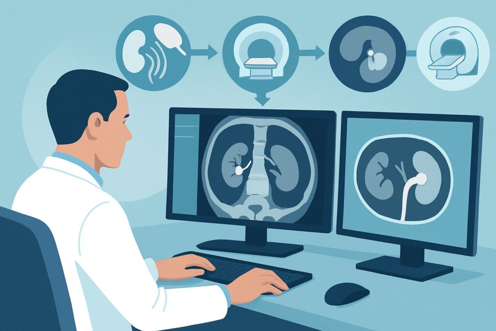
A 45-year-old male with a known 6 mm non-obstructing left renal stone, seen on a computed tomography (CT) scan three months ago, presents to the emergency department with severe, colicky left flank pain. His symptoms are identical to his prior episodes. You suspect the known stone is now passing, but you need to confirm its current location and assess for complications like obstruction. The clinical question is not if he has a stone, but what that stone is doing now. This article provides a deep dive into the American College of Radiology (ACR) Appropriateness Criteria for this specific follow-up scenario, where a `CT abdomen and pelvis without IV contrast` is rated Usually Appropriate and serves as the cornerstone of the diagnostic workflow.
Who Fits This Clinical Scenario?
This guidance is specifically for patients presenting with acute flank pain who meet two key criteria: a known, current urolithiasis diagnosis confirmed on recent imaging, and a recurrence of symptoms typical for stone passage. “Recent” generally implies imaging within the last 6 to 12 months, providing a reliable baseline for the stone’s size and location.
This workflow is designed to answer questions about stone migration, new obstruction, or changes in stone size. It is distinct from other common presentations that require different imaging strategies. This article does not apply to:
- Patients with a first-time presentation of flank pain: These individuals require initial diagnostic imaging to confirm the presence of a stone. This falls under a different ACR variant for initial workup.
- Pregnant patients: Due to radiation concerns for the fetus, the imaging algorithm for pregnant patients with suspected urolithiasis prioritizes non-ionizing modalities like ultrasound and MRI.
- Patients with atypical symptoms: If the patient presents with fever, rigors, leukocytosis, or signs of sepsis, the differential diagnosis broadens to include complicated urolithiasis (e.g., pyonephrosis, abscess). In these cases, a contrast-enhanced CT may be necessary to evaluate for infection and perinephric inflammation, a different clinical question.
What Diagnoses Are You Working Up in This Scenario?
When a patient with a known stone has recurrent symptoms, the imaging goal is to pinpoint the cause of the current acute episode. The differential diagnosis is focused and drives the choice of imaging modality.
Stone Migration with Ureteral Obstruction
This is the most common and clinically important diagnosis to confirm or exclude. The previously identified non-obstructing renal stone may have migrated into the ureter, causing partial or complete obstruction. The imaging study must accurately locate the stone within the ureter and quantify the degree of secondary hydronephrosis and perinephric stranding.
Passage of a Different, Unidentified Stone
In patients who are chronic stone formers, it’s possible that a smaller, previously unseen stone is the cause of the current symptoms, while the larger, known stone remains in the kidney. The imaging study should be sensitive enough to detect small calculi anywhere along the urinary tract.
Complicated Urolithiasis
While less common in patients without systemic signs of infection, an obstructing stone can lead to pyonephrosis (infected, obstructed kidney) or a perinephric abscess. Imaging helps identify secondary signs of inflammation or fluid collections that would necessitate urgent urologic intervention and antibiotic therapy.
Alternative or Coexisting Diagnosis
Though the pre-test probability for stone disease is high, the imaging study should be comprehensive enough to identify other potential causes of flank pain if no obstructing stone is found. Diagnoses like diverticulitis, appendicitis (if right-sided), or pyelonephritis can mimic stone pain and are readily identifiable on the recommended imaging study.
Why Is CT Abdomen and Pelvis without IV Contrast the Recommended Study for This Presentation?
The ACR designates `CT abdomen and pelvis without IV contrast` as Usually Appropriate for this scenario because it provides the most definitive answers to the key clinical questions with high speed and accuracy. Its diagnostic utility is unmatched for identifying the size, location, and downstream effects of a ureteral stone.
Non-contrast CT has exceptional sensitivity and specificity for detecting calcified stones of nearly all compositions and sizes. It precisely localizes the stone within the ureter (proximal, mid, or distal), which is critical information for urologic management planning. Furthermore, it clearly depicts secondary signs of obstruction, including hydroureter, hydronephrosis, and perinephric fat stranding, which correlate with the acuteness and severity of the obstruction.
Alternative studies are rated lower for specific reasons in this context:
- US kidneys and bladder retroperitoneal: Rated May be appropriate, this study is excellent for detecting hydronephrosis, which confirms an obstruction is present. However, it is notoriously poor at directly visualizing the stone itself, especially in the mid-ureter, due to overlying bowel gas. It is a valuable radiation-free option if the primary question is simply “is there obstruction?” but falls short when precise stone localization is needed for intervention planning.
- Radiography abdomen and pelvis (KUB): Also rated May be appropriate, a KUB is only useful for tracking the progression of a known, radiopaque (calcium-based) stone. It cannot detect radiolucent stones (e.g., uric acid), provides no information about obstruction (hydronephrosis), and cannot evaluate for alternative diagnoses. Its role is very limited in the acute setting.
A key consideration with CT is radiation exposure (adult relative radiation level ☢☢☢ 1-10 mSv). For recurrent stone formers who may require multiple scans over their lifetime, it is crucial to use low-dose CT protocols. These protocols significantly reduce the radiation dose while maintaining high diagnostic accuracy for stone detection. Intravenous contrast is not needed to visualize stones and is omitted to avoid unnecessary contrast exposure and to prevent the contrast from obscuring small calculi in the collecting system.
Once you’ve decided on CT abdomen and pelvis without IV contrast, our protocol guide covers the technique, contrast, and reading principles: CT Abdomen/Pelvis Without Contrast (Renal Stone).
What’s Next After CT Abdomen and Pelvis without IV Contrast? Downstream Workflow
The results of the non-contrast CT directly guide the subsequent clinical management. The workflow branches based on whether an obstructing stone is found.
If the study is positive for an obstructing ureteral stone:
Management depends on stone size, location, and the severity of symptoms. For small stones (<5 mm) in the distal ureter with manageable pain, medical expulsive therapy (e.g., tamsulosin) and observation are often appropriate. For larger stones (>10 mm), stones causing high-grade obstruction, or in cases of intractable pain or renal failure, an urgent urology consultation is required for intervention. This may involve ureteral stent placement, ureteroscopy with laser lithotripsy, or shock wave lithotripsy.
If the study is negative for an obstructing stone:
If the CT shows the known stone remains non-obstructing in the kidney and no other stone is seen, the patient’s pain may be due to non-obstructive renal colic or a passed stone. The focus shifts to pain management and medical therapy to prevent future stone growth. If pain persists or is atypical, further workup for non-urologic causes of flank pain should be considered based on findings from the CT scan (e.g., incidental appendicitis or diverticulitis).
If the study is indeterminate:
This is rare with modern CT for stone disease. However, if a ureteral filling defect is equivocal (e.g., differentiating a stone from a phlebolith or vessel crossing), a follow-up CT with IV contrast (CT Urogram) may be considered. This scenario routes to a different ACR variant: “Acute onset flank pain. Suspicion of stone disease. CT without contrast is inconclusive for the presence of stones.”
Pitfalls to Avoid (and When to Get Help)
In this specific scenario, several common pitfalls can lead to diagnostic delays or unnecessary radiation exposure. Be mindful of the following:
- Defaulting to contrast: Ordering a CT “with contrast” by default is a frequent error. IV contrast can obscure small stones within the collecting system and is not necessary for the primary diagnosis.
- Ignoring cumulative radiation dose: For young patients or known recurrent stone formers, failing to use a low-dose CT protocol contributes to a significant lifetime radiation burden. Always specify “low-dose” or “renal stone protocol” when ordering.
- Over-reliance on KUB: Using a plain radiograph to “rule out” a stone is inadequate. It will miss radiolucent stones and provides no information on the degree of obstruction.
- Misinterpreting phleboliths: Pelvic phleboliths (calcified veins) are a common mimic of distal ureteral stones. Look for the “comet tail sign” of a vessel extending from the calcification and trace the ureter carefully on all slices.
If the patient has signs of infection (fever, leukocytosis) in the setting of a confirmed obstructing stone, this constitutes a urologic emergency. Escalate immediately to a urology specialist for consideration of urgent decompression.
Related ACR Topics and Tools
Navigating imaging decisions requires access to reliable, evidence-based resources. For this scenario and related clinical questions, the following tools and guides are essential for ensuring appropriate care.
- For breadth across all scenarios in Acute Onset Flank Pain-Suspicion of Stone Disease (Urolithiasis), see our parent guide: Acute Onset Flank Pain-Suspicion of Stone Disease (Urolithiasis): ACR Appropriateness Decoded.
- ACR Appropriateness Criteria Lookup — for adjacent scenarios and different clinical presentations.
- Imaging Protocol Library — for detailed technical specifications on the recommended study.
- Radiation Dose Calculator — for discussing cumulative dose with patients who have recurrent disease.
Frequently Asked Questions
If my patient had a CT a year ago, is that ‘recent’ enough to use this follow-up workflow?
Generally, imaging within 6-12 months is considered recent enough to establish a baseline. If the prior imaging is older than a year, or if the patient’s clinical status has significantly changed, it may be more appropriate to treat the presentation as an initial episode of flank pain, although non-contrast CT remains the preferred study in most non-pregnant adults.
Why not just get an ultrasound first to avoid radiation?
Ultrasound is rated ‘May be appropriate’ and is a reasonable first step, especially in younger patients or if the main clinical question is simply confirming the presence of hydronephrosis. However, it often fails to visualize the stone itself, particularly in the mid-ureter. If definitive stone location and size are needed to plan a urologic intervention, a low-dose non-contrast CT is superior and often unavoidable.
The patient’s creatinine is elevated. Can I still order a non-contrast CT?
Yes. Since this is a CT scan performed *without* intravenous contrast, there is no risk of contrast-induced nephropathy. The patient’s renal function does not preclude the use of a non-contrast CT scan.
What if the CT shows the known stone hasn’t moved, but the patient is still in severe pain?
This finding suggests the pain may be from non-obstructive renal colic, the passage of a small stone that was missed or has already passed, or a non-urologic cause. The CT scan is still valuable as it can help rule out other intra-abdominal pathologies like diverticulitis or appendicitis. Management would then focus on pain control and metabolic workup to prevent future stone events.
Is a KUB (radiography) useful for follow-up after the CT?
Yes, once a radiopaque stone has been identified and located on CT, a KUB can be a very effective, low-radiation method to track its movement down the ureter or confirm its passage after a course of medical expulsive therapy. It is not, however, an adequate substitute for CT in the initial acute evaluation.
Reviewed by Pouyan Golshani, MD, Interventional Radiologist — May 30, 2026