What’s the Right First Imaging Step for Chronic Cough with Lung Cancer Risk?
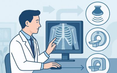
It’s a busy Tuesday afternoon in your primary care clinic. Your next patient is a 68-year-old man with a 45-pack-year smoking history who presents with a persistent, dry cough that has lingered for nearly three months. He has tried over-the-counter remedies without relief and is now worried. Given his significant risk factor for malignancy, you know that simply treating empirically for common causes of cough isn’t sufficient. The immediate clinical question is which imaging study to order first to evaluate for a serious underlying pathology without exposing him to unnecessary radiation or cost. According to the American College of Radiology (ACR) Appropriateness Criteria, for this specific presentation, a standard chest radiograph is rated Usually appropriate as the initial imaging step.
Who Fits This Clinical Scenario?
This guidance applies specifically to adult patients presenting with a chronic cough, defined as one lasting more than eight weeks, who also have an increased risk for lung cancer. The “increased risk” is a clinical determination based on well-established factors. Key inclusion criteria for this scenario include:
- A significant smoking history (current or former).
- A history of other tobacco use.
- Occupational exposures to known carcinogens like asbestos, radon, or silica.
- A personal history of prior cancers, particularly those with a tendency to metastasize to the lungs.
- A strong family history of lung cancer.
- Age over 50-55, particularly in combination with other risk factors.
It is crucial to distinguish this patient from those in closely related but distinct clinical situations. This workflow does not apply to patients with a chronic cough but no known risk factors for lung cancer; their workup may follow a different path. Similarly, this guidance is for initial imaging. It does not apply to a patient who has already undergone a clinical evaluation, a course of empiric treatment (e.g., for asthma or gastroesophageal reflux disease), and continues to have persistent symptoms, which represents a separate ACR scenario requiring a different approach.
What Diagnoses Are You Working Up in This Scenario?
When ordering initial imaging for a high-risk patient with chronic cough, the primary goal is to rule out serious thoracic pathology. The differential diagnosis is broad, but the imaging is focused on identifying structural abnormalities.
Lung Cancer (Malignancy) is the most pressing concern and the principal driver for this specific ACR variant. A new or growing pulmonary nodule, a mass, mediastinal or hilar lymphadenopathy, or a post-obstructive pneumonia can all present with a persistent cough. The initial radiograph is a sensitive first-pass tool for detecting these overt findings.
Chronic Obstructive Pulmonary Disease (COPD) is highly prevalent in the same patient population (i.e., long-term smokers). While the diagnosis is primarily based on spirometry, imaging can reveal signs like hyperinflation, flattened diaphragms, or bullous changes. The cough may be a symptom of the underlying COPD itself.
Interstitial Lung Disease (ILD) encompasses a group of disorders characterized by inflammation and/or scarring of the lung tissue. Patients may present with a dry, persistent cough and shortness of breath. A chest radiograph can reveal characteristic patterns like reticulation or honeycombing, prompting further investigation with high-resolution CT.
Chronic Infection, while less common in many communities, remains an important consideration. Tuberculosis, particularly post-primary or reactivation disease, can present with chronic cough and is often visible on a chest radiograph as apical opacities or cavities. Fungal infections can also cause chronic pulmonary symptoms and radiographic changes.
Why Is a Chest Radiograph the Recommended Initial Study?
For a patient with chronic cough and lung cancer risk factors, the American College of Radiology designates a standard two-view (PA and lateral) Radiography chest as Usually appropriate. This recommendation is based on a careful balance of diagnostic yield, safety, and resource utilization for an initial evaluation.
A chest radiograph serves as an excellent first-line screening tool. It is highly effective at detecting significant parenchymal, pleural, and mediastinal abnormalities that would warrant immediate further action. It can readily identify suspicious masses, large nodules, significant lymphadenopathy, pleural effusions, and consolidations suggestive of post-obstructive pneumonia. Its ability to provide a global assessment of the thorax makes it the ideal starting point.
Furthermore, it is a low-cost, widely accessible examination with a very low radiation dose. The ACR notes the relative radiation level (RRL) as ☢ (<0.1 mSv), which is a fraction of the dose from other cross-sectional imaging modalities and is comparable to the background radiation a person receives naturally over a few days.
Why are other studies rated lower for this initial step?
- CT chest without or with IV contrast is rated as May be appropriate. While CT is far more sensitive than radiography for detecting small nodules and subtle interstitial changes, it is not the recommended first test. Its significantly higher radiation dose (☢☢☢ 1-10 mSv) and greater cost make it better suited as a problem-solving tool after an abnormal or equivocal chest radiograph, or if clinical suspicion remains extremely high despite a negative radiograph.
- FDG-PET/CT skull base to mid-thigh is rated as Usually not appropriate for this initial workup. A PET/CT is a functional imaging study used primarily for cancer staging, not for the initial diagnosis of an undifferentiated cough. Its use at this stage would be premature, exposing the patient to a very high radiation dose (☢☢☢☢ 10-30 mSv) without a confirmed malignancy to evaluate.
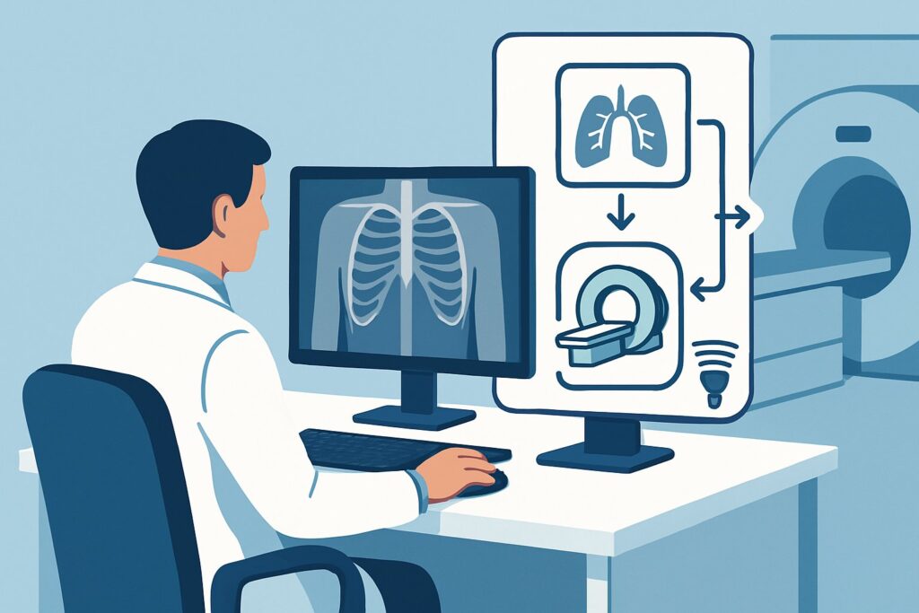
What’s Next After a Chest Radiograph? Downstream Workflow
The results of the initial chest radiograph will dictate the subsequent clinical pathway. The workflow is a decision tree based on whether the findings are positive, negative, or indeterminate.
If the radiograph is positive for a suspicious finding (e.g., a non-calcified nodule, a mass, hilar adenopathy, or a persistent opacity), the next step is nearly always a CT chest. A CT with intravenous contrast is often preferred to better characterize the lesion, assess for vascular involvement, and evaluate the mediastinum and hila for lymphadenopathy. This finding would also trigger an urgent referral to a pulmonologist or thoracic oncologist for further management, which may include biopsy via bronchoscopy or percutaneous needle aspiration.
If the radiograph is negative and shows no acute or suspicious process, the focus shifts back to the common, non-malignant causes of chronic cough. The patient should be evaluated for asthma, gastroesophageal reflux disease (GERD), and upper airway cough syndrome (postnasal drip). This may involve empiric treatment trials, spirometry, or referral to an allergist or gastroenterologist. A negative radiograph in a high-risk patient is reassuring but does not completely exclude a small, early-stage cancer. If the cough persists despite treatment for these common causes, the patient may then fit the criteria for a different ACR scenario: “Chronic cough lasting more than 8 weeks. Persistent symptoms despite initial clinical evaluation and empiric treatment,” where a CT chest may become appropriate.
If the radiograph is indeterminate (e.g., shows a small, ill-defined opacity or subtle interstitial changes), a follow-up CT chest is often warranted to provide a more definitive characterization.
Pitfalls to Avoid (and When to Get Help)
In this specific clinical workflow, several common pitfalls can delay diagnosis or lead to unnecessary testing. First, avoid “satisfaction of search”—if you see evidence of COPD, do not stop looking for a discrete nodule that could represent a concurrent malignancy. Second, do not dismiss a negative chest radiograph as definitive if high clinical suspicion persists; a small, central, endobronchial tumor may not be visible on a plain film. Third, ensure you order both PA and lateral views, as the lateral view is critical for evaluating the retrosternal and retrocardiac spaces. If the cough is accompanied by red-flag symptoms like hemoptysis, significant unintentional weight loss, or new-onset hoarseness, consider escalating directly to a CT scan and making an urgent referral to pulmonology, even if the initial radiograph is pending or negative.
Related ACR Topics and Tools
This article focuses on one specific clinical variant. For a comprehensive overview of imaging for all common chronic cough presentations, from low-risk patients to those with persistent symptoms after treatment, please see our parent guide. For additional resources to help select the right test and understand the technical details, the following GigHz tools are available.
- For breadth across all scenarios in Chronic Cough, see our parent guide: Chronic Cough: ACR Appropriateness Decoded.
- ACR Appropriateness Criteria Lookup — For exploring adjacent scenarios or different clinical questions.
- Imaging Protocol Library — For technical details on how specific studies are performed.
- Radiation Dose Calculator — For discussing cumulative radiation exposure with patients.
Frequently Asked Questions
If my patient is a smoker but under 50, does this guidance still apply?
Yes, this guidance is based on risk factors, not a strict age cutoff. A significant smoking history (e.g., 20+ pack-years) confers increased risk for lung cancer regardless of age. The clinical suspicion might be lower in a younger patient, but the initial imaging choice of a chest radiograph remains the most appropriate first step.
Why not just start with a low-dose CT (LDCT) like in lung cancer screening?
Lung cancer screening with LDCT is for asymptomatic, high-risk individuals meeting specific criteria (age 50-80, ≥20 pack-year history, current or former smoker within 15 years). This patient is symptomatic with a chronic cough. While a CT is more sensitive, the ACR recommends starting with a chest radiograph for an initial diagnostic workup of a symptom, reserving CT for follow-up of an abnormal radiograph or persistent symptoms.
What if the chest radiograph is negative but the cough gets worse or changes?
A change in the character of a chronic cough (e.g., new sputum production, new hemoptysis) or worsening severity despite a negative initial radiograph is a significant clinical development. This warrants prompt re-evaluation and often justifies proceeding to a CT chest to look for an underlying cause that may have been too subtle for the radiograph to detect.
Does a history of asbestos exposure change the initial imaging recommendation?
No, a history of significant asbestos exposure is a major risk factor for both lung cancer and mesothelioma, placing the patient squarely in this clinical scenario. A chest radiograph remains the appropriate initial imaging test. It is effective at identifying pleural plaques, effusions, or parenchymal changes (asbestosis) that would warrant a follow-up CT for better characterization.
If the patient has hemoptysis in addition to a chronic cough, should I still start with a chest radiograph?
Hemoptysis is a red-flag symptom that significantly increases the pre-test probability of serious pathology like lung cancer or bronchiectasis. While a chest radiograph is still a reasonable first step and rated ‘Usually Appropriate’ in the ACR criteria for hemoptysis, many clinicians may choose to proceed directly to a CT chest with contrast, which is also rated ‘Usually Appropriate’ for that indication. The decision depends on the severity of hemoptysis and overall clinical context.
Reviewed by Pouyan Golshani, MD, Interventional Radiologist — May 29, 2026