What’s the Right First Imaging Test for Suspected Biliary Disease with RUQ Pain?
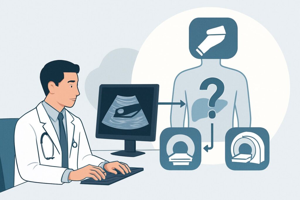
A 48-year-old woman presents to the emergency department with 4 hours of severe, colicky right upper quadrant pain that began after a fatty dinner. The pain radiates to her right shoulder, and she reports nausea. Her vital signs are stable, and while she is tender in the right upper quadrant, there are no peritoneal signs. Her labs are pending, but your clinical suspicion is high for biliary disease, likely acute cholecystitis. You need to choose the best initial imaging study to confirm the diagnosis and guide management. This is the classic decision point this article addresses.
For this specific scenario—right upper quadrant pain with suspected biliary disease—the American College of Radiology (ACR) Appropriateness Criteria rate US abdomen as Usually Appropriate, making it the clear first-choice examination.
Who Fits This Clinical Scenario?
This guidance is for patients presenting with signs and symptoms classic for a primary biliary pathology. The typical presentation involves acute-onset, severe, and persistent right upper quadrant (RUQ) or epigastric pain. The pain is often described as colicky, may radiate to the back or right shoulder (Boas’ sign), and is frequently associated with nausea, vomiting, and exacerbation after a fatty meal. This patient profile strongly suggests conditions like cholelithiasis (gallstones), acute cholecystitis, or choledocholithiasis (common bile duct stones).
This workflow is specifically for the initial imaging evaluation. It assumes no prior relevant imaging has been performed for this episode of pain. It’s crucial to distinguish this scenario from similar but distinct clinical situations that require a different approach:
- Unknown Etiology: If the pain is atypical or the differential is broad beyond the biliary tree (e.g., suspected liver abscess, renal colic, or perforated ulcer), the scenario of RUQ Pain, Unknown Etiology may be more fitting, potentially altering the choice of initial imaging.
- Negative or Equivocal Ultrasound: If an ultrasound has already been performed and was negative or inconclusive, but clinical suspicion for biliary disease remains high, you have advanced to a different clinical problem. The next imaging choice is addressed in scenarios like RUQ pain, suspected biliary disease, negative or equivocal ultrasound.
- Suspected Acalculous Cholecystitis: In critically ill, septic, or postoperative patients, acalculous cholecystitis is a key consideration. While ultrasound is still often the first step, the downstream workflow and alternative studies differ significantly.
What Diagnoses Are You Working Up in This Scenario?
When ordering the initial imaging for suspected biliary disease, the primary goal is to identify or exclude a few key, high-prevalence conditions. The differential diagnosis guides the radiologist’s search pattern and helps you interpret the clinical significance of the findings.
Acute Cholecystitis: This is the most urgent diagnosis to confirm or rule out. It is most commonly caused by a gallstone obstructing the cystic duct, leading to gallbladder inflammation, edema, and potentially ischemia or perforation. Imaging looks for the causative stone as well as secondary signs like gallbladder wall thickening (>3 mm), pericholecystic fluid, and a sonographic Murphy’s sign (maximal tenderness over the sonographically-located gallbladder).
Cholelithiasis (Symptomatic Gallstones): Even without acute inflammation, gallstones can cause significant pain, known as biliary colic. Ultrasound is exceptionally sensitive for detecting stones within the gallbladder lumen. Identifying stones confirms a diagnosis and provides a target for eventual surgical intervention if symptoms are recurrent.
Choledocholithiasis: This refers to a gallstone that has migrated into the common bile duct (CBD), which can cause obstruction, jaundice, and potentially life-threatening ascending cholangitis. While ultrasound can sometimes visualize a stone in the CBD, its most important role is often detecting indirect evidence, such as biliary ductal dilatation, which strongly suggests a downstream obstruction and prompts further investigation with studies like MRCP.
Less Common Considerations: The initial ultrasound can also reveal other pathologies mimicking biliary pain, such as a liver abscess, hepatic mass, or complications like gallbladder polyps or sludge. While not the primary targets, their identification is a key benefit of the initial sonographic evaluation.
Why Is US abdomen the Recommended Initial Study for This Presentation?
The ACR designates an abdominal ultrasound (US) as “Usually Appropriate” for the initial evaluation of suspected biliary disease because it excels in answering the most critical clinical questions in this scenario with high diagnostic accuracy and no patient risk from radiation.
The primary strength of ultrasound is its high sensitivity and specificity for detecting cholelithiasis. It can identify gallstones as small as 1-2 mm and is considered the gold standard for this purpose. For acute cholecystitis, its diagnostic power is enhanced by the ability to perform a real-time dynamic assessment, including testing for the sonographic Murphy’s sign. This combination of anatomic and physiologic information is unique to ultrasound.
Furthermore, ultrasound carries an adult relative radiation level of O (0 mSv), making it unequivocally safe for all patients, including pregnant women and younger individuals for whom cumulative radiation exposure is a concern. It is widely available, relatively inexpensive, and can often be performed at the bedside in unstable patients.
Why are alternative studies rated lower for initial imaging?
- CT abdomen with IV contrast is rated “May be appropriate.” While excellent for evaluating complications like perforation or abscess, CT is less sensitive than ultrasound for detecting gallstones, as many stones are isodense to bile and can be missed. It also involves significant ionizing radiation (☢☢☢ 1-10 mSv) and the risks of IV contrast. It is better reserved for when the diagnosis is unclear after ultrasound or when complications are suspected.
- MRI abdomen with MRCP is also rated “May be appropriate.” Magnetic resonance cholangiopancreatography (MRCP) is the best non-invasive test for evaluating the biliary ducts and detecting choledocholithiasis. However, it is more expensive, less available, and takes longer to perform than ultrasound. It is not a cost-effective or efficient first-line tool for diagnosing simple cholecystitis but serves as an outstanding problem-solving study when ultrasound is equivocal or shows ductal dilatation.
- Nuclear medicine scan (HIDA) is rated “May be appropriate.” A HIDA scan is a functional study that assesses the patency of the cystic duct. It is highly sensitive and specific for acute cholecystitis but provides no anatomic information about gallstones or other structures. It is typically used as a secondary test when ultrasound results are equivocal.
What’s Next After US abdomen? Downstream Workflow
The results of the initial abdominal ultrasound will direct your next steps. The clinical pathway branches significantly based on whether the findings are positive, negative, or indeterminate.
If the study is positive for acute cholecystitis or symptomatic cholelithiasis: A definitive diagnosis has been made. The next step is a surgical consultation for consideration of cholecystectomy. The timing of surgery (urgent vs. elective) will depend on the severity of the inflammation and the patient’s overall clinical status.
If the study is negative (no stones, no wall thickening, no ductal dilatation): A negative ultrasound makes acute calculous cholecystitis highly unlikely. At this point, you must reconsider your differential diagnosis. If clinical suspicion for a biliary cause remains high (e.g., classic symptoms, abnormal liver function tests), the patient now fits a different clinical scenario: Right upper quadrant pain, suspected biliary disease, negative or equivocal ultrasound. The next appropriate step in that workflow may be a functional study like a HIDA scan to evaluate for biliary dyskinesia or acalculous cholecystitis, or an MRCP if there is concern for a small, missed common bile duct stone.
If the study is indeterminate or equivocal: This is a common and challenging outcome. Examples include a thickened gallbladder wall without stones, or a dilated common bile duct without a visible obstructing stone. In the case of a dilated CBD, the next logical step is an MRI with MRCP, which is rated “May be appropriate” and is the best non-invasive test to visualize the entire biliary tree and identify a potential stone, stricture, or mass. If the gallbladder itself is abnormal but the findings are non-specific, a HIDA scan may be used to confirm or exclude cystic duct obstruction.
Pitfalls to Avoid (and When to Get Help)
While ultrasound is a robust tool, several clinical and technical pitfalls can lead to diagnostic errors in the workup of RUQ pain.
- Ignoring Patient Fasting Status: A patient who has recently eaten will have a physiologically contracted gallbladder. This state makes it extremely difficult to assess the gallbladder wall, search for small stones, or elicit a reliable sonographic Murphy’s sign, potentially leading to a false-negative study. Ensure patients are NPO for at least 6-8 hours if possible.
- Overlooking Acalculous Cholecystitis: In the right clinical setting (e.g., a critically ill ICU patient), the absence of stones does not rule out cholecystitis. Maintain a high index of suspicion for acalculous cholecystitis if the ultrasound shows secondary signs of inflammation.
- Body Habitus Limitations: In patients with obesity or significant bowel gas, sonographic windows can be poor, limiting visualization of the gallbladder and biliary ducts. The report should comment on the quality of the study; a limited study may need to be followed by CT or MRI if suspicion remains high.
- Misinterpreting Ductal Dilation: While a dilated common bile duct (typically >6-7 mm) is a red flag for obstruction, it can also be a normal finding in patients post-cholecystectomy. Always correlate with the patient’s surgical history.
If a patient shows clinical signs of ascending cholangitis (Charcot’s triad: fever, jaundice, RUQ pain) or sepsis, this is a medical emergency. Escalate immediately for surgical and/or gastroenterology consultation for potential urgent biliary decompression via ERCP, regardless of the initial imaging findings.
Related ACR Topics and Tools
This article covers one specific variant within the broader topic of Right Upper Quadrant Pain. For a comprehensive overview of all related scenarios and their recommended imaging pathways, please consult our parent guide. You can also use the tools below to explore other ACR criteria, imaging techniques, and radiation safety considerations.
- For breadth across all scenarios in Right Upper Quadrant Pain, see our parent guide: Right Upper Quadrant Pain: ACR Appropriateness Decoded.
- To explore other clinical scenarios, use the ACR Appropriateness Criteria Lookup.
- For details on imaging techniques, see the Imaging Protocol Library.
- To discuss radiation exposure with patients, use the Radiation Dose Calculator.
Frequently Asked Questions
Why not just get a CT scan on every patient with RUQ pain in the emergency department?
While CT is a powerful tool, it is not the best initial test for suspected biliary disease. Ultrasound is more sensitive for detecting gallstones and does not use ionizing radiation. A CT scan exposes the patient to a significant radiation dose (1-10 mSv) and is best reserved for cases where the diagnosis is unclear after ultrasound or when a complication like a perforation or abscess is suspected.
What should I do if the patient just ate before they need an ultrasound?
A recent meal causes the gallbladder to contract, which can hide gallstones and prevent accurate assessment of the gallbladder wall, potentially leading to a false-negative result. If the situation is not emergent, the scan should be delayed until the patient has been fasting for at least 6 hours. If the scan must be done emergently, it’s critical to communicate the patient’s post-prandial state to the radiologist, as a ‘negative’ result may be unreliable.
Does a negative ultrasound definitively rule out a gallbladder problem?
No. A negative ultrasound is excellent for ruling out gallstones and typical signs of acute cholecystitis. However, it cannot rule out functional disorders like biliary dyskinesia (a problem with gallbladder emptying) or acalculous cholecystitis (inflammation without stones). If clinical suspicion remains high despite a negative ultrasound, a HIDA scan may be the appropriate next step to assess gallbladder function.
When is it appropriate to order an MRCP instead of an ultrasound as the first test?
It is rarely appropriate to order an MRCP as the very first test for undifferentiated RUQ pain. Its primary role is as a second-line or problem-solving study. The main indication for an MRCP is when there is clinical or sonographic suspicion of a common bile duct stone (choledocholithiasis), such as jaundice, elevated bilirubin, or a dilated bile duct seen on ultrasound. In these cases, MRCP is the best non-invasive way to confirm or exclude a ductal stone.
Is a HIDA scan the same as a gallbladder ultrasound?
No, they are fundamentally different tests. An ultrasound uses sound waves to create an anatomical image of the gallbladder and surrounding structures, looking for stones and signs of inflammation. A HIDA scan (hepatobiliary iminodiacetic acid scan) is a nuclear medicine functional study. It involves injecting a radiotracer that is taken up by the liver and excreted into the bile, allowing visualization of bile flow. Its primary use is to determine if the cystic duct is blocked, which is the hallmark of acute cholecystitis, or to measure the gallbladder ejection fraction to diagnose biliary dyskinesia.
Reviewed by Pouyan Golshani, MD, Interventional Radiologist — June 27, 2026