When to Order Imaging for Infective Endocarditis: ACR Appropriateness Decoded
When to Order Imaging for Infective Endocarditis: ACR Appropriateness Decoded
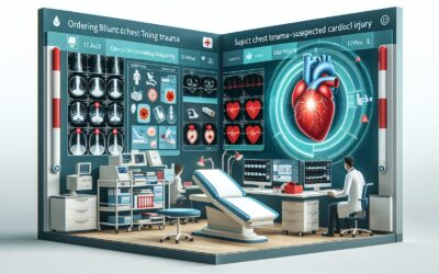
It’s late in the evening, and you’re evaluating a patient with a new-onset heart murmur, fever, and a history of intravenous drug use. Blood cultures are pending, but your clinical suspicion for infective endocarditis (IE) is high. The immediate question is which imaging study to order first. Do you start with a transthoracic echocardiogram (TTE), or is the clinical picture concerning enough to consider a transesophageal echocardiogram (TEE) or a cardiac CT? Choosing the right initial and follow-up imaging is critical for diagnosis, risk stratification, and guiding treatment. This guide decodes the American College of Radiology (ACR) Appropriateness Criteria to help you make evidence-based decisions for your patients with suspected IE.
What Does the ACR Guideline for Infective Endocarditis Cover?
The ACR Appropriateness Criteria for Infective Endocarditis focus on the diagnostic imaging pathway for patients where this condition is a primary consideration. The guideline is structured around two common clinical scenarios: the initial workup of a patient with suspected IE, and the subsequent imaging needed to guide management or treatment once the diagnosis is known or strongly suspected. It provides a framework for selecting the most suitable modality based on diagnostic yield, invasiveness, and radiation exposure.
This guideline specifically addresses the evaluation of native and prosthetic heart valves for vegetations, abscesses, and other complications of endocarditis. It does not cover routine screening in asymptomatic patients, the evaluation of non-cardiac sources of infection, or imaging for septic emboli to other organ systems, which are addressed in separate ACR guidelines. The recommendations are intended for both adult and pediatric populations, with specific attention to radiation safety in younger patients.
What Imaging Should I Order for Infective Endocarditis? Recommendations by Clinical Scenario
The ACR panel provides clear, scenario-based recommendations to guide imaging selection for infective endocarditis. The optimal study depends on whether you are performing an initial evaluation or require additional detail for patient management.
For a patient with suspected infective endocarditis undergoing initial imaging, a transthoracic echocardiogram (TTE) is rated as Usually appropriate. This non-invasive ultrasound is the cornerstone of initial evaluation, providing excellent visualization of valvular morphology and function to detect vegetations. A chest radiograph is also Usually appropriate to assess for concomitant pulmonary findings like septic emboli or heart failure. A dedicated CT of the heart with IV contrast is also considered Usually appropriate, offering high-resolution anatomical detail, particularly for evaluating perivalvular complications like abscesses, which may be difficult to see on TTE. A transesophageal echocardiogram (TEE) is rated as May be appropriate (Disagreement) for initial imaging; while it offers superior sensitivity for small vegetations and prosthetic valve endocarditis, its semi-invasive nature often positions it as a second-line or problem-solving tool after an inconclusive TTE.
For a patient with known or suspected infective endocarditis requiring additional imaging to direct patient management or treatment, the recommendations shift. In this context, a transesophageal echocardiogram (TEE) is rated as Usually appropriate. TEE is invaluable for pre-operative planning, confirming the extent of perivalvular extension, and assessing for complications like leaflet perforation or abscess formation. TTE remains Usually appropriate for follow-up and monitoring treatment response. A CT of the heart with IV contrast is also Usually appropriate in this setting, serving as a powerful alternative or adjunct to TEE, especially in patients who cannot tolerate TEE or when evaluating prosthetic valve dehiscence. Advanced modalities like FDG-PET/CT heart and cardiac MRI with and without contrast are rated as May be appropriate, typically reserved for complex cases, such as suspected prosthetic valve endocarditis with equivocal echo findings or when assessing myocardial involvement.
ACR Imaging Recommendations Table for Infective Endocarditis
| Clinical Scenario | Top Procedure | ACR Rating | Adult RRL | Pediatric RRL |
|---|---|---|---|---|
| Suspected infective endocarditis. Initial imaging. | US echocardiography transthoracic resting | Usually appropriate | O 0 mSv | O 0 mSv [ped] |
| Known or suspected infective endocarditis. Additional imaging to direct patient management or treatment. | US echocardiography transesophageal | Usually appropriate | O 0 mSv | O 0 mSv [ped] |
Adult vs. Pediatric Infective Endocarditis Imaging: Radiation Dose Tradeoffs
When imaging children for suspected infective endocarditis, the principle of As Low As Reasonably Achievable (ALARA) is paramount. The ACR guidelines reflect this by emphasizing non-ionizing radiation modalities first. Both transthoracic and transesophageal echocardiography are radiation-free and are the primary imaging tools in both adult and pediatric populations. However, when CT is necessary, the radiation dose (RRL) levels are tiered differently. For example, a CT of the heart with IV contrast carries a high dose in adults (☢ ☢ ☢ ☢ 10-30 mSv) but is categorized in a lower dose tier for children (☢ ☢ ☢ ☢ 3-10 mSv), reflecting the use of size- and weight-based protocols to minimize exposure.
This distinction is critical due to the increased radiosensitivity of developing tissues and the longer lifespan over which the risks of radiation-induced malignancy can manifest. Clinicians must weigh the diagnostic necessity of a CT scan against the cumulative radiation burden for the pediatric patient. In cases where CT is required to assess for complex complications like abscesses or pseudoaneurysms, using pediatric-specific low-dose protocols is essential.
Imaging Protocol Details for Infective Endocarditis
Once you’ve decided on the right study based on the ACR criteria, ensuring it is performed correctly is the next critical step. The diagnostic quality of a cardiac CT or echocardiogram depends heavily on the specific imaging protocol, including contrast timing, ECG gating, and acquisition parameters. Standardized protocols help ensure that the images acquired will be sufficient to answer the clinical question and avoid the need for repeat studies. While this article focuses on which study to order, detailed procedural guides are available to help your team execute the scan properly.
Tools to Help You Order the Right Study
Navigating imaging guidelines and patient-specific factors can be complex. GigHz offers a suite of reference tools designed to support clinical decision-making at the point of care.
For scenarios beyond infective endocarditis, the ACR Appropriateness Criteria Lookup provides a searchable interface to the full library of ACR guidelines, helping you find evidence-based recommendations for hundreds of clinical variants.
To ensure studies are performed to the highest standard, the Imaging Protocol Library offers detailed, step-by-step protocols for a wide range of CT, MRI, and ultrasound examinations, promoting consistency and quality.
When discussing the risks and benefits of imaging with patients, especially when ionizing radiation is involved, the Radiation Dose Calculator is a valuable resource for estimating cumulative exposure and communicating this information in an understandable way.
Is a TTE or TEE better for diagnosing infective endocarditis?
A transthoracic echocardiogram (TTE) is the recommended first-line imaging test because it is non-invasive, widely available, and has good specificity. However, a transesophageal echocardiogram (TEE) is more sensitive, especially for detecting small vegetations (<5 mm), evaluating prosthetic valves, and identifying perivalvular complications like abscesses. The ACR rates TTE as “Usually appropriate” for initial imaging, while TEE is often used as a follow-up test if the TTE is negative or inconclusive but clinical suspicion remains high.
When is a cardiac CT indicated for infective endocarditis?
A cardiac CT with IV contrast is rated “Usually appropriate” for both initial and follow-up imaging. It is particularly valuable for assessing perivalvular and extracardiac complications, such as abscesses, pseudoaneurysms, and fistulae. It can be an excellent alternative or complementary study to TEE, especially in post-operative patients or those who cannot tolerate a TEE. ECG-gated cardiac CT provides detailed anatomical information that can be critical for surgical planning.
What is the role of FDG-PET/CT in infective endocarditis?
FDG-PET/CT is rated “May be appropriate” for additional imaging to direct management, particularly in complex cases. Its primary role is in diagnosing prosthetic valve endocarditis, where metallic artifact can limit echocardiography and CT. The increased metabolic activity (FDG uptake) in infected tissue can help confirm the diagnosis when other imaging is equivocal. It is not recommended for the initial workup of native valve endocarditis.
Why is cardiac MRI usually not appropriate for initial evaluation?
For the initial evaluation of suspected infective endocarditis, cardiac MRI (with or without contrast) is rated “Usually not appropriate.” While MRI provides excellent soft tissue characterization, it is less sensitive than echocardiography for detecting small, mobile vegetations. Its longer acquisition time and limited availability make it less practical for acute diagnosis compared to TTE or TEE. MRI’s role is rated as “May be appropriate” for problem-solving in later stages, such as evaluating myocardial involvement or characterizing abscesses, but it is not a first-line tool.
Should I order a chest radiograph for suspected endocarditis?
Yes, a chest radiograph is rated “Usually appropriate” for the initial imaging of a patient with suspected infective endocarditis. While it cannot diagnose endocarditis directly, it is crucial for evaluating for related cardiopulmonary complications, such as septic pulmonary emboli (common in right-sided endocarditis), pulmonary edema from valvular regurgitation, or cardiomegaly.
Reviewed by Pouyan Golshani, MD, Interventional Radiologist — May 12, 2026