Which Imaging Is Best for a High-Risk Stress Fracture After a Negative Radiograph?
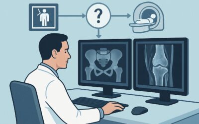
A 68-year-old woman with osteoporosis, on long-term bisphosphonate therapy, presents to your clinic with three weeks of worsening, localized pain in her right thigh. She denies any specific trauma, but the pain is sharp with weight-bearing. You obtain radiographs of her femur, which show no acute fracture or other obvious abnormality. Given her risk profile for an atypical femoral fracture, the clinical suspicion for an occult insufficiency fracture remains high, and the possibility of fracture completion is a serious concern. You need to decide on the next, more sensitive imaging study to confirm or exclude the diagnosis and guide immediate management. This article provides a clinical workflow for this exact scenario, where the American College of Radiology (ACR) rates an MRI of the area of interest without IV contrast as Usually Appropriate.
Who Fits This Clinical Scenario?
This guidance is for an adult patient with a suspected stress fracture (either fatigue or insufficiency type) where there is a high risk of the fracture completing or an immediate need to establish a definitive diagnosis. The key inclusion criteria are:
- An adult patient.
- Clinical suspicion for a stress fracture in any location except the vertebrae.
- Initial radiographs are negative, equivocal, or indeterminate.
- The patient is considered high-risk for fracture completion or progression. This includes individuals with known osteoporosis, those on long-term bisphosphonate therapy (risk for atypical femoral fractures), patients with other metabolic bone diseases, or elite athletes with suspected high-risk fractures (e.g., femoral neck, navicular).
This workflow is distinct from other similar presentations. This article does not apply if you are ordering the initial imaging for a suspected stress fracture, as radiographs are typically the first step. It also does not apply to pregnant patients, who have unique considerations for radiation and imaging timing. Finally, this guidance is for diagnosis; if a stress fracture has already been confirmed on radiographs and the goal is surgical planning, a different set of imaging considerations applies.
What Diagnoses Are You Working Up in This Scenario?
When ordering advanced imaging for a high-risk patient with focal bone pain and negative radiographs, you are evaluating a specific differential diagnosis where a missed or delayed diagnosis could lead to significant morbidity.
Impending or Occult Insufficiency Fracture: This is the primary concern, especially in an older patient with metabolic bone disease. In patients on long-term bisphosphonates, this includes the particularly concerning atypical femoral fracture, which often presents with prodromal thigh or groin pain. These fractures begin as a stress reaction on the lateral cortex and can progress to a complete, transverse fracture with minimal trauma.
High-Grade Bone Stress Reaction: This represents a precursor to a true fracture. Pathophysiologically, it involves significant bone marrow edema and periosteal reaction from repetitive mechanical stress that has overwhelmed the bone’s ability to repair itself. Detecting this early allows for intervention (e.g., non-weight-bearing) to prevent progression to a full fracture.
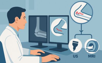
Musculotendinous Injury: Soft tissue injuries can mimic the pain of a stress fracture. For example, deep gluteal pain can be caused by a sacral stress fracture or by proximal hamstring tendinopathy. An imaging study should be able to differentiate between bone and soft tissue pathology.
Underlying Malignancy: Though less common, a primary bone tumor (like osteosarcoma) or a metastatic lesion can present as focal bone pain and weaken the bone, leading to a pathologic fracture. These are critical “can’t-miss” diagnoses that advanced imaging can help identify.
Osteomyelitis: A bone infection can also cause focal pain and may not be apparent on initial radiographs. While less likely without specific risk factors like diabetes, recent surgery, or immunocompromise, it remains in the differential for unexplained, severe focal bone pain.
Why Is MRI Without IV Contrast the Recommended Study for This Presentation?
For a high-risk patient with negative radiographs, Magnetic Resonance Imaging (MRI) of the area of interest without IV contrast is rated Usually Appropriate by the ACR. This recommendation is based on its superior diagnostic performance, safety profile, and ability to guide immediate clinical decisions in this specific context.
The primary strength of MRI is its exceptional sensitivity for detecting bone marrow edema. This edema is the earliest sign of a bone stress injury, appearing long before cortical changes or a visible fracture line develop. Fluid-sensitive sequences, such as Short Tau Inversion Recovery (STIR) or T2-weighted fat-suppressed images, vividly demonstrate this edema, allowing for diagnosis at the earliest, most treatable stage. Furthermore, MRI provides excellent anatomic detail, clearly defining the presence and extent of a fracture line if one has developed and assessing for signs of an impending atypical femoral fracture, such as lateral cortical thickening.
Alternative studies are rated lower for compelling reasons:
- CT of the area of interest without IV contrast is rated May be appropriate. While CT is superior to radiographs for visualizing subtle cortical breaks, it is insensitive to the early marrow edema that precedes a visible fracture. In a high-risk scenario where the goal is to intervene before fracture completion, waiting for a CT-visible fracture line may be too late.
- A whole-body bone scan with SPECT or SPECT/CT is rated May be appropriate (Disagreement). This nuclear medicine study is highly sensitive to areas of increased bone turnover. However, it is not specific; arthritis, infection, and tumors can all produce a “hot spot.” This lack of specificity can lead to diagnostic uncertainty. Critically, it also involves a significant radiation dose (adult_rrl=☢☢☢ 1-10 mSv), whereas MRI involves none (adult_rrl=O 0 mSv).
For this indication, IV contrast is deemed Usually not appropriate for the MRI. The key diagnostic findings of edema and fracture lines are well-visualized on non-contrast sequences. Adding contrast increases cost and carries a small risk of adverse reaction without providing significant additional information for differentiating a stress fracture from its most common mimics.
What’s Next After MRI? Downstream Workflow
The results of the MRI will directly dictate your next steps, which is why obtaining it promptly in a high-risk patient is so crucial. The downstream workflow typically follows one of three paths.
If the MRI is positive for a high-grade stress fracture or an incomplete atypical femoral fracture: This is an urgent clinical finding. The next step is immediate orthopedic surgery consultation. The patient should be placed on strict non-weight-bearing or toe-touch weight-bearing status for the affected limb to prevent fracture completion, which can occur with minimal force. Prophylactic surgical fixation is often required.
If the MRI is positive for a low-grade stress reaction (marrow edema without a discrete fracture line): Management is typically non-operative. This involves a period of protected weight-bearing (e.g., with crutches) and significant activity modification. The underlying cause, such as osteoporosis or training errors, should be addressed. Follow-up is typically managed by a primary care, sports medicine, or physical medicine and rehabilitation physician.
If the MRI is negative for any bone stress injury: This result effectively rules out a stress fracture and indicates the pain generator is likely elsewhere. The high-resolution soft tissue detail from the MRI may reveal an alternative diagnosis, such as tendinopathy, muscle tear, or bursitis. The next step is to treat the identified soft tissue pathology, often with physical therapy and activity modification. If the MRI is entirely negative, a broader re-evaluation of the differential diagnosis is warranted.
Pitfalls to Avoid (and When to Get Help)
In this high-stakes clinical scenario, several common pitfalls can delay diagnosis or lead to poor outcomes. First, avoid the “wait and see” approach. For a high-risk patient, repeating radiographs in 10-14 days (a strategy rated Usually not appropriate) consumes valuable time during which an incomplete fracture could displace. Second, do not order a vague study; be specific on the requisition (e.g., “MRI right femur to rule out atypical femoral fracture, pain localized to proximal third”). This ensures the radiologist uses the correct protocol focused on the area of concern. Finally, avoid ordering MRI with contrast, as it is unnecessary for this indication. If the MRI report describes a transverse line through the thickened lateral cortex of the subtrochanteric femur, escalate immediately to an orthopedic surgeon, as this is a classic sign of an impending atypical femoral fracture requiring urgent intervention.
Related ACR Topics and Tools
For a comprehensive overview of all clinical variants related to this topic, from initial imaging to post-operative evaluation, please consult the parent guide. For additional decision support and technical details, the following resources are available:
- For breadth across all scenarios in Stress (Fatigue/Insufficiency) Fracture, Including Sacrum, Excluding Other Vertebrae, see our parent guide: Stress (Fatigue/Insufficiency) Fracture, Including Sacrum, Excluding Other Vertebrae: ACR Appropriateness Decoded.
- To explore imaging recommendations for other clinical presentations, use the ACR Appropriateness Criteria Lookup.
- For detailed technical specifications of the recommended MRI, see the Imaging Protocol Library.
- To discuss radiation exposure from alternative studies like CT or bone scan with your patients, consult the Radiation Dose Calculator.
Frequently Asked Questions
Why not just repeat the radiograph in 10-14 days instead of ordering an MRI?
For a high-risk patient, waiting 10-14 days for a potential fracture line to become visible on a radiograph poses an unacceptable risk of the fracture completing in the interim. The ACR rates this approach as ‘Usually not appropriate’ for this specific scenario because the need for a definitive, timely diagnosis is high. MRI can detect the injury at its earliest stage.
What specifically makes a patient ‘high risk’ for fracture completion?
A patient is considered high risk if they have underlying conditions that compromise bone quality or are subject to extreme mechanical stress. This includes patients with osteoporosis, those on long-term bisphosphonate therapy (risk of atypical femoral fracture), individuals with other metabolic bone diseases, or high-performance athletes with pain in anatomically high-risk locations like the femoral neck or tarsal navicular.
Is a nuclear medicine bone scan a reasonable alternative if MRI is unavailable or contraindicated?
A bone scan is rated ‘May be appropriate (Disagreement)’ by the ACR. It can be used as a second-line option if MRI is not feasible. While it is very sensitive for detecting bone turnover, it is not specific—many conditions can cause a positive result. It also exposes the patient to a moderate amount of ionizing radiation (1-10 mSv), unlike MRI, which has none. MRI remains the strongly preferred study due to its superior specificity and safety.
Does this guidance apply to suspected high-risk stress fractures in the foot or ankle?
Yes. The clinical scenario excludes vertebrae but applies to the rest of the appendicular skeleton. The principles of using MRI for early diagnosis in high-risk situations are particularly relevant for locations in the foot and ankle known for poor healing or risk of nonunion, such as the tarsal navicular, talus, or the base of the fifth metatarsal.
Why is intravenous (IV) contrast not needed for this type of MRI?
The primary findings of a stress injury—bone marrow edema and a potential fracture line—are best visualized on non-contrast, fluid-sensitive MRI sequences (like STIR or T2 fat-suppressed) and T1-weighted sequences. IV contrast is typically used to evaluate for infection or tumor by assessing tissue vascularity. While these are on the differential, they are less likely, and contrast adds little value for the primary question of a stress fracture, while increasing cost and adding potential risk.
Reviewed by Pouyan Golshani, MD, Interventional Radiologist — May 30, 2026