Which Imaging Is Best for Chronic Cerebellar Ataxia in Adults? An ACR Workflow

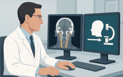
A 68-year-old patient presents to your neurology clinic with a six-month history of progressively worsening balance. They describe a feeling of unsteadiness, not true spinning vertigo, and their family has noted their gait is becoming wider and more clumsy. On examination, you confirm a broad-based gait, truncal instability, and clear dysmetria on finger-to-nose testing. You suspect a cerebellar process and must decide on the most appropriate initial imaging study. This article provides a clinical workflow for this exact scenario, detailing why the American College of Radiology (ACR) rates MRI head without and with IV contrast as Usually Appropriate.
Who Fits This Clinical Scenario?
This guidance applies specifically to adult patients presenting with a chronic and progressive or persistent sense of disequilibrium, where the physical examination reveals objective signs of cerebellar ataxia. Understanding these key components is crucial for applying this workflow correctly.
- Chronic: The symptoms have developed over weeks to months, or even years. This is not an acute or sudden-onset event.
- Disequilibrium: The patient’s primary complaint is a sense of imbalance or unsteadiness, particularly when standing or walking. This is distinct from vertigo, which is a sensation of spinning or movement.
- Cerebellar Ataxia: The diagnosis is supported by physical exam findings pointing to cerebellar dysfunction. These may include gait ataxia (wide-based, staggering walk), truncal ataxia (inability to sit or stand without swaying), limb ataxia (dysmetria, dysdiadochokinesia), or scanning speech.
This workflow is distinct from other similar presentations. It does not apply to:
- Acute Vertigo: Patients with a sudden onset of severe vertigo, which may be due to a peripheral cause (like vestibular neuritis) or a central cause (like a posterior circulation stroke). These scenarios have their own dedicated imaging pathways.
- Episodic, Positional Vertigo: Brief episodes of vertigo triggered by head movements are more suggestive of benign paroxysmal positional vertigo (BPPV), which typically does not require initial imaging.
- Sensory Ataxia: Patients whose imbalance is due to loss of proprioception (e.g., from dorsal column pathology or peripheral neuropathy). These patients often have a positive Romberg sign with eyes closed and lack the classic signs of cerebellar dysfunction like dysmetria.
What Diagnoses Are You Working Up in This Scenario?
In an adult with chronic cerebellar ataxia, the differential diagnosis is broad, spanning degenerative, structural, toxic, and metabolic causes. The initial imaging is designed to identify or exclude structural abnormalities that require specific management.
Cerebellar Degeneration and Atrophy
This is a primary consideration and includes a wide spectrum of disorders. Multiple system atrophy cerebellar type (MSA-C), spinocerebellar ataxias (SCAs), and idiopathic late-onset cerebellar ataxia (ILOCA) all cause progressive atrophy of the cerebellum and its connections. While imaging may be normal early on, it can later reveal characteristic patterns of volume loss in the cerebellum, pons, and middle cerebellar peduncles that help narrow the differential.
Posterior Fossa Structural Lesions
A slow-growing tumor is a critical, can’t-miss diagnosis. Primary brain tumors (e.g., hemangioblastoma) or, more commonly, metastatic disease to the cerebellum can present insidiously with progressive ataxia. Chronic subdural hematomas or arachnoid cysts can also exert mass effect on the cerebellum, leading to these symptoms.
Demyelinating Disease
Multiple Sclerosis (MS) can present with cerebellar signs, and a demyelinating plaque in the cerebellum or brainstem could be the underlying cause. While MS often presents earlier in life and with relapsing-remitting symptoms, a progressive form or an initial presentation with cerebellar signs is possible in adults.
Toxic or Metabolic Causes
While often a diagnosis of exclusion based on history, imaging can show the downstream effects of certain insults. The most common is alcohol-related cerebellar degeneration, which leads to characteristic atrophy of the superior cerebellar vermis. Chronic phenytoin use can also cause similar changes. Paraneoplastic cerebellar degeneration, an autoimmune process triggered by an occult malignancy, may show inflammatory changes or later-stage atrophy.
Why Is MRI Head Without and With IV Contrast the Recommended Study?
The ACR designates MRI head without and with IV contrast as Usually Appropriate for this clinical scenario because it provides the most comprehensive evaluation of the cerebellum and posterior fossa structures, which are the anatomical focus of the workup.
The superior soft-tissue resolution of Magnetic Resonance Imaging (MRI) is essential for detecting the subtle parenchymal changes associated with the key differential diagnoses. It can clearly visualize the cerebellar folia, deep cerebellar nuclei, and brainstem, allowing for the detection of:
- Atrophy: MRI is highly sensitive for volume loss, a key feature of degenerative cerebellar disorders like MSA-C or SCAs.
- Tumors: Both primary and metastatic tumors in the posterior fossa are well-visualized on MRI. The addition of intravenous (IV) gadolinium-based contrast is critical, as many tumors demonstrate characteristic enhancement patterns that are essential for diagnosis.
- Demyelination: T2-weighted and FLAIR sequences are highly sensitive for the demyelinating plaques of MS, which appear as high-signal lesions. Active inflammation will typically enhance with contrast.
- Ischemia: MRI can detect chronic lacunar infarcts or evidence of prior strokes that could contribute to the clinical picture.
The ACR also rates MRI head without IV contrast as Usually Appropriate. This may be a reasonable alternative in patients with severe contrast allergies or renal impairment, or when the pre-test probability for an enhancing lesion like a tumor or active MS is considered extremely low. However, the contrast-enhanced study provides a more complete initial assessment.
Why Other Studies Are Less Appropriate
Alternative imaging modalities are rated lower for this specific presentation for clear reasons:
- CT Head (Without or With Contrast): Rated Usually not appropriate. Computed Tomography (CT) is significantly limited in its ability to visualize the posterior fossa. Beam-hardening artifacts from the dense petrous temporal bones obscure fine details of the cerebellum and brainstem. CT will miss subtle atrophy, non-hemorrhagic strokes, small tumors, and demyelinating plaques.
- MRA Head and Neck: Rated Usually not appropriate. While Magnetic Resonance Angiography (MRA) is excellent for visualizing blood vessels, the primary concern in chronic, progressive ataxia is parenchymal disease, not a vascular emergency like an occlusion or aneurysm. It does not provide the detailed tissue characterization needed to evaluate for atrophy, tumors, or demyelination.
The recommended MRI study involves no ionizing radiation (0 mSv), which is a significant advantage over CT (1-10 mSv).
What’s Next After MRI Head Without and With IV Contrast? Downstream Workflow
The results of the MRI will guide the subsequent diagnostic and management pathway. The workflow branches based on whether the findings are positive, negative, or indeterminate.
If the MRI is positive for a structural lesion:
- Tumor or Mass: An urgent referral to neurosurgery and/or neuro-oncology is warranted for biopsy and treatment planning. A full systemic workup for a primary cancer may be needed if metastasis is suspected.
- Demyelinating Plaques: If findings are suggestive of Multiple Sclerosis, the next steps typically include an MRI of the cervical and thoracic spine to look for additional lesions and a lumbar puncture to test for oligoclonal bands in the cerebrospinal fluid.
- Evidence of Stroke: Management would focus on secondary stroke prevention, including management of vascular risk factors.
If the MRI shows a specific atrophy pattern:
- Findings like pontocerebellar atrophy (suggesting MSA-C) or isolated vermian atrophy (suggesting alcohol-related degeneration) can help confirm a clinical diagnosis. The next steps involve neurology follow-up for symptom management, and in some cases, genetic testing for hereditary ataxias (SCAs).
If the MRI is negative or non-specific:
- A normal MRI is a very common and important result, as it effectively rules out major structural causes. The workup then pivots away from structural imaging to focus on non-structural causes. This includes comprehensive laboratory testing for vitamin deficiencies (B12, E, thiamine), antibody testing for paraneoplastic syndromes and gluten ataxia, and potentially genetic testing.
Pitfalls to Avoid (and When to Get Help)
Navigating the workup for chronic ataxia requires careful consideration to avoid common missteps.
- Ordering CT First: A frequent error is ordering a non-contrast CT of the head as a “screening” test. Due to its poor sensitivity for posterior fossa pathology, a negative CT provides false reassurance and merely delays the definitive study, which is an MRI.
- Omitting Contrast: Forgoing IV contrast without a compelling contraindication may cause you to miss an enhancing tumor or an active demyelinating plaque, which are critical diagnoses to make.
- Misinterpreting Mild Atrophy: Age-related generalized volume loss is common. Attributing a patient’s significant ataxia solely to “mild age-related cerebellar atrophy” can prematurely end the search for other treatable metabolic, toxic, or autoimmune causes.
- Ignoring the Spine: If the clinical picture is ambiguous between cerebellar and sensory ataxia, remember that proprioceptive pathways run through the spinal cord. In such cases, an MRI of the spine may be warranted if the brain MRI is unrevealing. The ACR rates MRI cervical and thoracic spine without IV contrast as May be appropriate for this reason.
If the diagnosis remains elusive after initial imaging and laboratory workup, referral to a neurologist with expertise in movement disorders or ataxia is the appropriate next step.
Related ACR Topics and Tools
For a comprehensive overview of imaging for all types of imbalance, from acute vertigo to chronic disequilibrium, please consult our parent topic guide. For tools to help select and understand imaging studies, see the resources below.
- For breadth across all scenarios in Dizziness and Ataxia, see our parent guide: Dizziness and Ataxia: ACR Appropriateness Decoded.
- ACR Appropriateness Criteria Lookup — for adjacent scenarios
- Imaging Protocol Library — for technique on the recommended study
- Radiation Dose Calculator — for cumulative dose conversations
Frequently Asked Questions
Is an MRI without contrast sufficient for initial imaging of chronic cerebellar ataxia?
According to the ACR, an MRI of the head without IV contrast is also rated ‘Usually Appropriate’. It can effectively assess for atrophy, chronic strokes, and established demyelinating plaques. However, an MRI with contrast is more comprehensive as it is superior for detecting enhancing tumors or active inflammatory lesions, which are critical diagnoses to exclude. The choice may depend on patient factors and pre-test probability, but the combined study is generally preferred for a complete initial workup.
My patient has a pacemaker. Can I still order an MRI for their ataxia?
Many modern pacemakers and implantable cardiac devices are MRI-conditional. However, this requires a specific protocol and close collaboration with the radiology department and cardiology. The patient’s device must be checked for MRI compatibility, and a specific protocol must be followed before, during, and after the scan. If the patient’s device is an absolute contraindication to MRI, a high-resolution CT with contrast may be the next best option, despite its limitations in the posterior fossa.
If the brain MRI is normal, what is the next step in the workup?
A normal brain MRI is a crucial finding that rules out most structural causes like tumors, strokes, or significant demyelination. The workup should then shift to non-structural causes. This typically involves blood work to test for vitamin deficiencies (B1, B12, E), thyroid function, and screening for celiac disease (anti-gliadin/anti-transglutaminase antibodies). Testing for paraneoplastic antibodies and genetic testing for spinocerebellar ataxias (SCAs) may also be considered based on the clinical context and family history.
Why is MRA not recommended for this specific scenario?
Magnetic Resonance Angiography (MRA) is designed to visualize blood vessels, not the brain parenchyma. In the setting of chronic, progressive cerebellar ataxia, the primary concern is a disease process within the brain tissue itself (e.g., degeneration, tumor, inflammation). While rare vascular malformations could be a cause, they are not the primary target of the initial investigation. Therefore, a standard parenchymal MRI is the appropriate first-line study, and MRA is rated ‘Usually not appropriate’ for this indication.
What if my patient’s ataxia is accompanied by signs of a peripheral neuropathy?
If there are signs of both cerebellar ataxia and sensory deficits (e.g., loss of vibration or position sense), you may be dealing with a process that affects both the central and peripheral nervous systems. This is seen in conditions like Friedreich’s ataxia, vitamin E deficiency, or certain paraneoplastic syndromes. While the initial brain MRI is still the correct first step, an MRI of the spine may also be considered to evaluate the dorsal columns. The ACR rates ‘MRI cervical and thoracic spine without IV contrast’ as ‘May be appropriate’ in this scenario.
Reviewed by Pouyan Golshani, MD, Interventional Radiologist — May 30, 2026