Which Imaging Is Best for Liver Cancer After Local Therapy? An ACR-Guided Workflow
A 68-year-old man with cirrhosis and a 4 cm hepatocellular carcinoma (HCC) in segment VI underwent transarterial chemoembolization (TACE) three months ago. He returns to your oncology clinic for follow-up, reporting stable health. His alpha-fetoprotein (AFP) level has decreased but not fully normalized. You now face a critical decision: how to accurately assess the tumor’s response to treatment to determine if he needs further therapy, can proceed to surveillance, or is a candidate for transplant. This article details the American College of Radiology (ACR) Appropriateness Criteria workflow for this specific scenario: post-treatment evaluation of primary liver cancer. For this presentation, the ACR rates MRI abdomen without and with IV contrast as Usually Appropriate.
Who Fits This Clinical Scenario for Post-Treatment Liver Cancer Evaluation?
This guidance applies to adult patients with a known diagnosis of primary liver cancer, most commonly hepatocellular carcinoma (HCC), who have completed a course of liver-directed therapy or neoadjuvant chemotherapy. The goal of imaging is to evaluate the therapeutic response, not for initial diagnosis or routine screening.
Inclusion Criteria:
- Adult patient with confirmed primary liver cancer (e.g., HCC, intrahepatic cholangiocarcinoma).
- Recent completion of locoregional therapy such as transarterial chemoembolization (TACE), radiofrequency ablation (RFA), stereotactic body radiation therapy (SBRT), or radioembolization (Y-90).
- Recent completion of neoadjuvant chemotherapy or systemic therapy prior to planned resection or transplant.
This workflow is distinct from other related clinical situations. This guidance does not apply to:
- Initial Staging: Patients with a newly diagnosed liver mass who have not yet received treatment. This requires a different imaging approach focused on characterizing the lesion and detecting metastatic disease.
- Screening: High-risk patients (e.g., with cirrhosis) undergoing imaging to detect a new liver cancer.
- Routine Surveillance: Patients who have already demonstrated a complete response to treatment and are now in a long-term follow-up protocol.
Correctly identifying the clinical scenario is the first step to selecting the most appropriate and highest-value imaging study.
What Diagnoses Are You Working Up After Liver-Directed Therapy?
In the post-treatment setting, the “differential diagnosis” shifts from identifying the type of cancer to assessing the treatment effect. The primary goal is to distinguish viable, active tumor from successfully treated, non-viable tissue. The key possibilities the imaging study aims to differentiate are:
Complete Response
This is the ideal outcome, defined by the absence of any viable tumor tissue within the treated area. Imaging should show no signs of arterial phase hyperenhancement (APHE) or other features of active cancer. This finding moves the patient into a surveillance pathway.
Partial Response or Residual Viable Tumor
This is the most critical finding to identify. It means that while the therapy was effective to some degree, a portion of the tumor remains active and requires further treatment. Imaging will demonstrate persistent, often nodular or irregular, areas of enhancement characteristic of viable HCC.
Progressive Disease
This indicates that the tumor has grown despite treatment, or new intrahepatic or extrahepatic lesions have appeared. This is a significant finding that necessitates a change in management, often to a different local therapy or systemic treatment.
Treatment-Related Changes
Locoregional therapies induce changes in the liver parenchyma, such as inflammation, necrosis, fibrosis, and hemorrhage. These changes can sometimes mimic residual tumor, creating a diagnostic challenge. A high-quality imaging study is essential to differentiate benign post-treatment enhancement from malignant tissue.
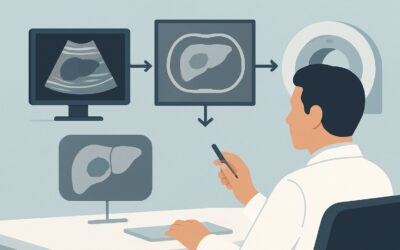
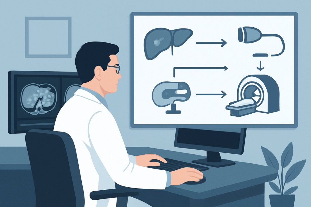
Why Is MRI of the Abdomen the Recommended Study for Post-Treatment Evaluation?
The ACR designates MRI abdomen without and with IV contrast as a Usually Appropriate procedure for evaluating treatment response in primary liver cancer. Its superior soft-tissue contrast resolution is the primary reason for this recommendation, as it allows for a more confident distinction between viable tumor and post-treatment changes.
The key to assessing HCC viability is identifying its hallmark vascular pattern: hyperenhancement in the late arterial phase with subsequent “washout” of contrast in the portal venous or delayed phases. MRI is exceptionally sensitive for detecting this pattern. Furthermore, advanced techniques like diffusion-weighted imaging (DWI) can provide complementary information on tissue cellularity, and subtraction imaging is crucial for differentiating true contrast enhancement from the intrinsic high signal of TACE material or hemorrhage.
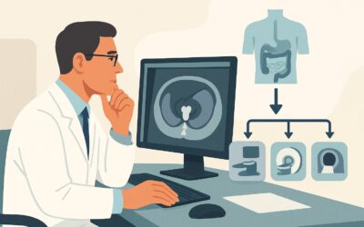
While MRI is the top-rated study, other modalities are also considered:
- CT abdomen with IV contrast multiphase is also rated Usually Appropriate. It is an excellent alternative, particularly if MRI is contraindicated (e.g., incompatible implanted devices, severe claustrophobia) or less accessible. Multiphase CT can effectively demonstrate the vascular patterns of HCC. However, it involves significant ionizing radiation (☢☢☢☢ 10-30 mSv), a key consideration for patients who will require serial follow-up scans. MRI’s superior ability to characterize subtle areas of enhancement often makes it the preferred initial choice.
- US abdomen with IV contrast is rated Usually Not Appropriate. While contrast-enhanced ultrasound (CEUS) can assess vascularity, it is highly operator-dependent, has a limited field of view, and is less effective at evaluating the entire liver, detecting new lesions, or assessing for extrahepatic disease compared to MRI or CT.
When ordering the recommended MRI, it is critical to specify a “multiphase liver protocol.” This ensures the radiology department acquires the specific pre-contrast, late arterial, portal venous, and delayed phase images necessary for a complete and accurate assessment based on the Liver Imaging Reporting and Data System (LI-RADS) criteria for treatment response.
What’s Next After the MRI? Interpreting Results and Planning the Next Step
The MRI report, ideally structured using the LI-RADS Treatment Response criteria, will guide your downstream clinical workflow. The findings will categorize the treated lesion as non-viable, viable, or equivocal.
If the study shows Complete Response (LR-TR Non-viable):
This is an excellent prognostic sign. The patient can transition to a routine surveillance protocol. The next imaging study will be part of the “Treated. Routine surveillance” scenario, typically performed at regular intervals (e.g., every 3-6 months) to monitor for local recurrence or new primary tumors.
If the study shows Residual or Recurrent Disease (LR-TR Viable):
The presence of viable tumor necessitates further action. The case should be discussed at a multidisciplinary tumor board to determine the best next step. Options may include:
- Repeat locoregional therapy (e.g., another round of TACE or ablation).
- Consideration for salvage therapies like SBRT.
- Evaluation for systemic therapy if the disease is extensive or locoregional options are exhausted.
- Re-evaluation of transplant candidacy.
If the study is Indeterminate (LR-TR Equivocal):
Sometimes, post-treatment inflammation and early recurrence can be difficult to distinguish. For an equivocal finding, the most common next step is a short-interval follow-up MRI, typically in 1 to 3 months, to assess for change. A growing or more conspicuous area of enhancement on the follow-up scan would confirm viable disease.
Common Pitfalls in Post-Treatment Liver Imaging (and When to Escalate)
Navigating post-treatment imaging requires careful attention to detail to avoid misinterpretation that could lead to under- or over-treatment.
- Misinterpreting Post-Ablation Rim Enhancement: A thin, smooth, uniform rim of enhancement around an ablation zone is a common and expected finding representing benign inflammatory tissue. In contrast, thick, nodular, or irregular enhancement along the margin is highly suspicious for residual or recurrent tumor.
- Using a Single-Phase Scan: Ordering a generic “CT with contrast” or “MRI with contrast” is a major pitfall. Without the specific multiphase protocol (especially the late arterial phase), the hallmark vascular features of viable HCC will be missed, rendering the study non-diagnostic for this indication.
- Ignoring Subtraction Images: In patients post-TACE, the retained high-density ethiodized oil can mimic enhancement on CT, and blood products can be bright on pre-contrast T1-weighted MRI. Subtraction imaging, which digitally removes the pre-contrast signal, is essential to confirm true enhancement.
If the imaging report suggests new vascular invasion, new extrahepatic metastases, or a rapidly progressing tumor burden, this represents a significant change in the patient’s status. Escalate immediately by presenting the case at a multidisciplinary tumor board to facilitate a prompt change in the treatment plan.
Related ACR Topics and Tools
This article covers one specific workflow within the broader topic of liver cancer imaging. For a comprehensive overview of all related scenarios, from screening to staging and surveillance, please consult our parent guide. The following GigHz tools can also support your clinical decision-making:
- Parent Topic Hub: For breadth across all scenarios in Staging and Follow-up of Primary Liver Cancer, see our parent guide: Staging and Follow-up of Primary Liver Cancer: ACR Appropriateness Decoded.
- ACR Criteria Lookup: To explore adjacent scenarios or search other clinical guidelines, visit the ACR Appropriateness Criteria Lookup.
- Protocol Library: For detailed technical parameters of the recommended study, see the Imaging Protocol Library.
- Dose Calculator: To discuss cumulative radiation exposure with patients considering CT, use the Radiation Dose Calculator.
Frequently Asked Questions
Why is MRI preferred over multiphase CT if both are rated ‘Usually Appropriate’?
While both are excellent tools, MRI is often preferred for two main reasons. First, it has superior soft tissue contrast, which can make it easier to distinguish subtle viable tumor from benign post-treatment inflammation or fibrosis. Second, MRI does not use ionizing radiation, which is a significant advantage for cancer patients who often require numerous follow-up scans over many years.
How soon after therapy should this imaging be performed?
The timing of the first post-treatment scan is crucial. Typically, it is performed 1 to 3 months after the procedure. Imaging too early can be misleading, as post-treatment inflammation can mimic residual disease. Imaging too late may allow for significant tumor progression if the treatment was not successful. The exact timing should be coordinated with the treating interventional radiologist or oncologist.
What if my patient has a contraindication to MRI, such as a non-compatible pacemaker?
In cases where MRI is contraindicated, multiphase CT of the abdomen is the appropriate alternative. It is also rated ‘Usually Appropriate’ by the ACR for this indication and provides the necessary information on tumor vascularity to assess treatment response. Be sure to order a ‘multiphase liver protocol’ to ensure the correct contrast timing.
Is a PET/CT scan useful for evaluating treatment response in HCC?
For this specific scenario, FDG-PET/CT is rated ‘Usually Not Appropriate’ by the ACR. While some poorly differentiated hepatocellular carcinomas are FDG-avid, many well-differentiated tumors are not, leading to a high rate of false-negative results. Furthermore, post-treatment inflammation is often FDG-avid, which can cause false-positive results. Therefore, multiphase MRI or CT remains the standard of care.
What is the role of the tumor marker AFP in conjunction with imaging?
Alpha-fetoprotein (AFP) is a valuable adjunct to imaging but should not be used alone to determine treatment response. A rising AFP after treatment is highly suspicious for residual or recurrent disease, even with equivocal imaging findings. Conversely, a normal or decreasing AFP does not rule out viable tumor. The imaging findings and AFP trend should always be interpreted together.
Reviewed by Pouyan Golshani, MD, Interventional Radiologist — May 30, 2026