Which Imaging Is Best for New Deficits in a Known Demyelinating Disease?
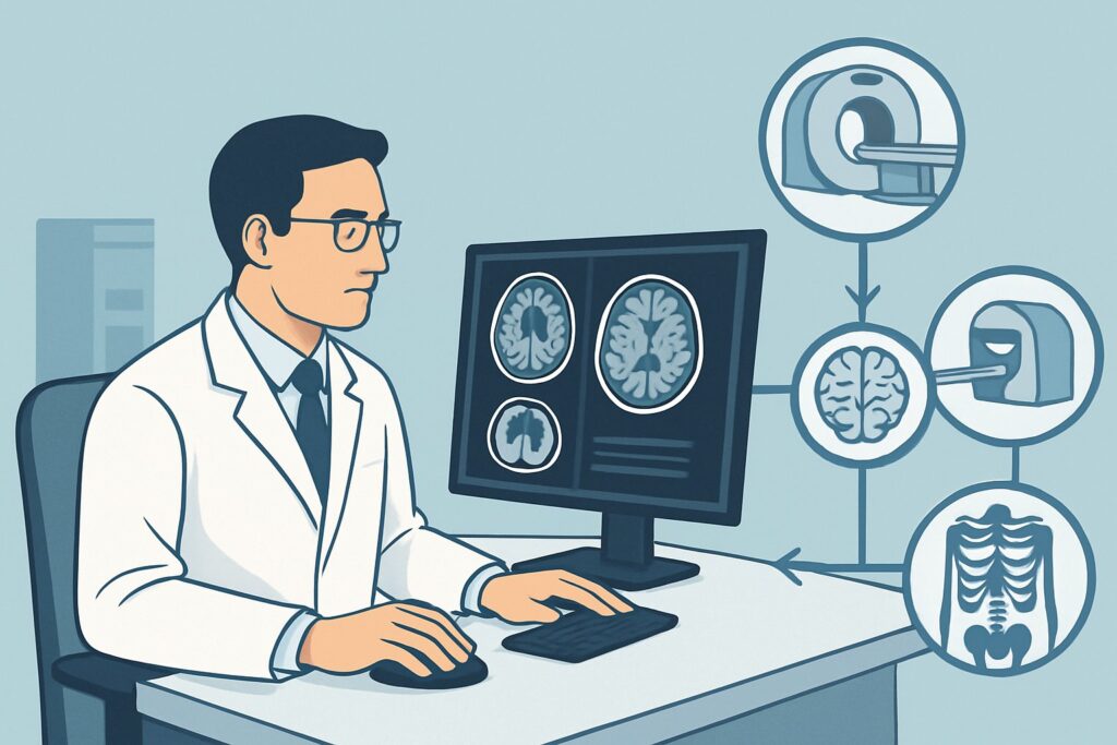
A 42-year-old woman with a known diagnosis of multiple sclerosis (MS) presents to your clinic with a two-week history of progressive leg weakness and a new sensory level at her mid-chest. She is worried this is a major relapse, and you need to determine the best initial imaging study to confirm disease activity and guide treatment changes. This scenario—an adult with a known demyelinating disease experiencing new or worsening neurologic deficits—requires a specific imaging strategy to differentiate a relapse from disease progression or an alternative diagnosis. According to the American College of Radiology (ACR) Appropriateness Criteria, the initial imaging study that is Usually Appropriate is an MRI of the cervical and thoracic spine without and with IV contrast. This article provides a detailed workflow for this exact clinical decision.
Who Fits This Clinical Scenario?
This guidance is specifically for an adult patient with an established diagnosis of a central nervous system (CNS) demyelinating disease, such as multiple sclerosis (MS), neuromyelitis optica spectrum disorder (NMOSD), or myelin oligodendrocyte glycoprotein antibody-associated disease (MOGAD). The key trigger for imaging is the presentation with new or clearly progressive neurologic symptoms—for example, new-onset weakness, a distinct sensory level, or worsening ataxia that represents a significant change from their baseline.
This workflow is distinct from several similar but separate clinical situations:
- First-time presentation: If a patient presents with initial symptoms suspicious for a demyelinating disease but has no prior diagnosis, they fit a different scenario. The workup is for a suspected new diagnosis, not a change in a known one.
- Stable surveillance: If a patient with known MS is neurologically stable and the imaging is for routine monitoring of disease burden or treatment response, that falls under the surveillance imaging scenario, which has different recommendations.
- Acute transverse myelitis suspicion: If the presentation is acutely suggestive of transverse myelitis with symptoms clearly localizing to a single spinal cord level, a more focused workup for that specific condition may be warranted.
Applying this article’s guidance is appropriate only when the pre-test probability of a relapse or significant progression in a known demyelinating condition is high.
What Diagnoses Are You Working Up in This Scenario?
When a patient with a known demyelinating disease presents with new deficits, the primary goal of imaging is to confirm the cause and rule out mimics. The differential diagnosis guides the choice of imaging modality and protocol.
Disease Relapse (New Inflammatory Lesion)
This is the most common and expected cause. A relapse is defined by new or worsening symptoms corresponding to a new area of active inflammation in the CNS. Imaging is crucial to objectively confirm this activity, typically identified as a new gadolinium-enhancing lesion on MRI. Confirming a relapse is essential for initiating acute treatment, such as high-dose corticosteroids, and considering a change in the patient’s long-term disease-modifying therapy (DMT).
Progressive Multifocal Leukoencephalopathy (PML)
A less common but critical diagnosis to consider, PML is a severe, often fatal, demyelinating disease caused by the reactivation of the John Cunningham (JC) virus. It is a known risk associated with certain DMTs (e.g., natalizumab). Its imaging appearance can sometimes mimic an MS relapse, but there are often distinguishing features a radiologist can identify. Given its severe prognosis, excluding PML is a high priority in the right clinical context.
Spinal Cord Compression or Other Structural Lesion
Not all new neurologic deficits in a patient with MS are caused by MS. A structural mimic, such as a herniated intervertebral disc, spinal stenosis, or a spinal cord tumor, can present with similar symptoms of myelopathy. MRI is excellent at identifying these alternative causes, which would lead to a completely different management pathway, such as a neurosurgical consultation.
Re-evaluation of Diagnosis (e.g., NMOSD vs. MS)
The presentation of a new spinal cord lesion can sometimes prompt a re-evaluation of the underlying diagnosis. For instance, the discovery of a longitudinally extensive transverse myelitis (LETM)—a lesion spanning three or more vertebral segments—is a hallmark of NMOSD or MOGAD and is atypical for MS. Identifying this finding would trigger a different serologic workup and has major implications for long-term treatment.
Why Is MRI of the Cervical and Thoracic Spine the Recommended Initial Study?
For an adult with a known demyelinating disease presenting with new deficits suggestive of a spinal cord lesion, the ACR designates MRI of the cervical and thoracic spine without and with IV contrast as Usually Appropriate. This recommendation is based on the modality’s superior ability to visualize the specific pathology in question without exposing the patient to ionizing radiation.
The rationale for this specific study is multi-faceted:
- High Sensitivity for Demyelination: MRI is unparalleled in its ability to detect demyelinating plaques within the spinal cord. T2-weighted and STIR (Short Tau Inversion Recovery) sequences are highly sensitive for identifying areas of edema and demyelination, appearing as bright signals within the cord. This allows for visualization of both new and old lesions.
- Assessment of Disease Activity: The administration of intravenous gadolinium-based contrast is essential. Active inflammatory lesions demonstrate breakdown of the blood-brain barrier, resulting in enhancement on post-contrast T1-weighted images. The presence of enhancement confirms a clinically suspected relapse and provides objective evidence of ongoing disease activity.
- Exclusion of Mimics: The same MRI study is the gold standard for ruling out structural causes of myelopathy, such as disc herniation, spinal stenosis, tumors, or vascular malformations.
Alternative studies are rated lower for clear reasons:
- CT of the Cervical and Thoracic Spine (Usually Not Appropriate): CT offers poor soft-tissue contrast and cannot reliably visualize demyelinating plaques within the spinal cord parenchyma. It is the wrong tool for the primary clinical question. Furthermore, it involves a significant radiation dose (☢☢☢☢ 10-30 mSv).
- MRI of the Lumbar Spine (Usually Not Appropriate): The spinal cord typically terminates at the L1-L2 vertebral level. In a patient with suspected myelopathy (upper motor neuron signs), imaging the lumbar spine, which contains the cauda equina (lower motor neurons), is a low-yield examination. The primary sites of involvement in MS are the cervical and thoracic cord.
Because MRI uses no ionizing radiation (0 mSv), it is the ideal modality for a patient population that may require multiple follow-up scans over their lifetime. Once you’ve decided on MRI of the spine, our protocol guide covers the foundational technique. For a deeper dive into the specific sequences and reading principles, see our guide: MRI Cervical Spine Without Contrast.
What’s Next After MRI? Downstream Workflow
The results of the spinal MRI will direct the subsequent clinical pathway. The workflow branches based on whether the findings are positive, negative, or indeterminate for active demyelination.
If the study is positive for a new enhancing lesion:
This result confirms an active disease relapse. The immediate next step is typically to initiate treatment with high-dose corticosteroids to reduce inflammation and hasten recovery. This finding should also prompt a discussion with the patient about the effectiveness of their current disease-modifying therapy and whether a change to a different agent is warranted.
If the study is negative for new or enhancing lesions:
A negative scan in the face of new symptoms can be diagnostically challenging. It may suggest that the symptoms are due to pseudo-relapse (e.g., triggered by infection or fever), functional neurologic symptoms, or a lesion located elsewhere in the CNS. The next step is often to order an MRI of the head without and with IV contrast, which is also rated Usually Appropriate for this scenario, to search for an active lesion in the brain or brainstem that could explain the symptoms.
If the study shows a non-demyelinating cause:
If the MRI reveals a structural mimic, such as significant spinal stenosis or a compressive disc herniation, the workflow shifts entirely. The next step is an urgent referral to a neurosurgeon or spine specialist for evaluation and management of the structural problem.
If the study is indeterminate or shows atypical features:
Findings like a longitudinally extensive lesion may require further workup to reconsider the primary diagnosis. This would involve serologic testing for AQP4 and MOG antibodies to rule out NMOSD and MOGAD, respectively. Consultation with a neuroradiologist or a neurologist specializing in demyelinating diseases can be invaluable in these complex cases.
Pitfalls to Avoid (and When to Get Help)
Navigating this clinical scenario requires avoiding several common pitfalls to ensure timely and accurate diagnosis.
- Ordering CT Instead of MRI: Never order a CT of the spine as the initial test to evaluate for an MS relapse. It lacks the sensitivity to answer the clinical question and exposes the patient to unnecessary radiation.
- Omitting IV Contrast: Ordering a non-contrast MRI to “save time” or avoid contrast is a critical error. Without gadolinium, you cannot distinguish active from chronic lesions, which is the central question in a suspected relapse.
- Imaging the Wrong Spinal Segment: Do not order an MRI of the lumbar spine for symptoms of myelopathy (e.g., spasticity, a thoracic sensory level). This is a low-yield study that will miss the pathology.
- Attributing All Symptoms to MS: Be careful not to have diagnostic fixation. Always consider structural mimics, especially if the clinical presentation is atypical for a relapse.
If the clinical picture and imaging findings are discordant, or if the diagnosis of PML is a possibility, escalate care by consulting with a neurologist who has expertise in demyelinating diseases.
Related ACR Topics and Tools
This article focuses on a single, specific clinical scenario. For a comprehensive overview of imaging across all presentations of demyelinating diseases, from initial suspicion to long-term surveillance, please refer to our parent guide. It provides a breadth of information that complements this deep-dive article.
- For breadth across all scenarios in Demyelinating Diseases, see our parent guide: Demyelinating Diseases: ACR Appropriateness Decoded.
- To explore other clinical scenarios, use the ACR Appropriateness Criteria Lookup.
- To review technical details for various studies, see the Imaging Protocol Library.
- For discussions about cumulative exposure with patients, use the Radiation Dose Calculator.
Frequently Asked Questions
Why is an MRI of the brain also rated ‘Usually Appropriate’ for this scenario?
An MRI of the head without and with IV contrast is also ‘Usually Appropriate’ because demyelinating lesions in the brain, brainstem, or cerebellum can cause a wide variety of neurologic deficits, including motor and sensory symptoms that might seem to originate from the spine. If spinal cord imaging is negative or the symptoms are ambiguous, brain imaging is the logical next step to locate a potential culprit lesion.
What if my patient has a contraindication to gadolinium contrast, like severe renal insufficiency?
If a patient cannot receive gadolinium, an MRI of the cervical and thoracic spine without IV contrast is rated as ‘May be appropriate’. While you lose the ability to definitively confirm lesion activity via enhancement, new or enlarging T2/STIR lesions can still strongly suggest a relapse. The decision to treat would then be based on a combination of the clinical presentation and these non-contrast imaging findings.
Should I order imaging of the entire spine (cervical, thoracic, and lumbar) at once?
No, this is generally not recommended. The ACR rates MRI of the lumbar spine as ‘Usually not appropriate’ for this indication because primary demyelinating plaques are very rare below the conus medullaris (L1-L2). Ordering a full spine MRI adds unnecessary time and cost for a very low diagnostic yield. The focus should be on the cervical and thoracic spine, where the vast majority of MS-related spinal cord lesions occur.
How soon after symptom onset should the MRI be performed?
Imaging should be performed as soon as is practical. While it is not typically a code-stroke level emergency, delaying the scan delays diagnosis and the initiation of treatment (like corticosteroids) that can speed recovery. Furthermore, gadolinium enhancement is a transient phenomenon, often lasting only 2-6 weeks, so performing the scan within that window is ideal to capture evidence of active inflammation.
Does a new, non-enhancing T2 lesion on MRI count as a relapse?
This is a nuanced clinical question. A new T2 lesion without enhancement indicates a new area of demyelination that is not acutely inflamed at the time of the scan. While it represents disease progression, whether it constitutes the ‘relapse’ causing the patient’s current symptoms is less certain. In this case, the decision to treat with steroids is more dependent on the severity of the clinical symptoms, as the imaging evidence for acute inflammation is absent.
Reviewed by Pouyan Golshani, MD, Interventional Radiologist — June 27, 2026