Which Imaging Is Best for Staging Late-Stage (IIB-III) ER+ Breast Cancer?
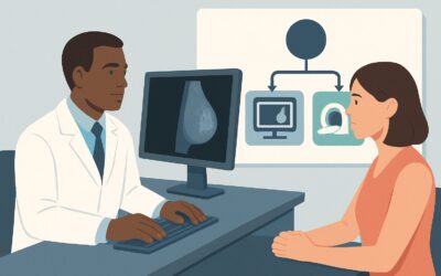
A 62-year-old woman presents to your oncology clinic with a new diagnosis of invasive ductal carcinoma. The biopsy confirms an estrogen receptor-positive (ER+), HER2-negative tumor. On examination, you palpate a 4 cm mass and firm, mobile axillary lymph nodes, placing her at clinical stage IIB at minimum. Before you can finalize a curative-intent treatment plan, you must first rule out distant metastatic disease. The critical question is which imaging studies are most appropriate to systemically stage her, evaluating the most common sites of spread: the bones, liver, and lungs. This article details the American College of Radiology (ACR) guided workflow for this specific scenario, where a Whole Body Bone Scan is rated Usually Appropriate as a foundational component of the workup.
Who Fits This Clinical Scenario for Breast Cancer Staging?
This guidance applies specifically to patients with a new diagnosis of invasive breast cancer—either invasive ductal carcinoma (IDC) or invasive lobular carcinoma (ILC)—that is ER-positive and HER2-negative. The key inclusion criterion is the clinical stage: this workflow is intended for patients presenting with late-stage, non-metastatic disease, defined as Stage IIB through Stage III. This stage is determined by the combination of tumor size (T) and lymph node involvement (N), typically involving larger tumors (T3, >5 cm) or significant axillary lymph node involvement (N1-N3).
It is crucial to distinguish this scenario from others that require a different approach:
- Early-Stage Disease (Stage I-IIA): Patients with smaller tumors and no clinical evidence of nodal spread have a low pre-test probability of distant metastases. Systemic staging is generally not recommended in this group, as the risk of false positives and unnecessary radiation exposure outweighs the potential benefit.
- Evaluation for Locoregional Disease: If the clinical question is to assess the extent of the tumor within the breast or to evaluate suspicious axillary nodes, the imaging workup is different. This often involves breast MRI or targeted axillary ultrasound, which are considered Usually Not Appropriate for the evaluation of distant disease.
- Aggressive Subtypes: While the staging modalities overlap, patients with triple-negative or HER2-positive breast cancer may benefit from FDG-PET/CT as a primary staging tool due to the higher metabolic activity and different metastatic patterns of these tumors.
What Diagnoses Are You Working Up in This Scenario?
For a patient with clinical stage IIB-III breast cancer, the primary goal of systemic imaging is to detect distant metastases. A positive finding upstages the patient to Stage IV, fundamentally shifting the treatment goal from curative to palliative and life-prolonging. The workup targets the most common sites of hematogenous spread for ER-positive breast cancer.
Bone Metastases
The skeleton is the most frequent site of distant disease for ER-positive breast cancer. Metastases can be asymptomatic or present with bone pain, and their detection is paramount for accurate staging. These lesions are typically osteoblastic (bone-forming), making them highly conspicuous on radionuclide bone scans which detect areas of increased bone turnover.
Visceral Metastases (Liver and Lungs)
The liver and lungs are the next most common sites of spread. Visceral metastases can significantly impact prognosis and treatment selection. Unlike bone metastases, these are not detectable on a bone scan and require cross-sectional imaging, such as a CT scan, for evaluation. Identifying these lesions is a co-primary goal of the staging workup.
Distant Nodal Metastases
Spread to lymph nodes beyond the axilla and internal mammary chain (e.g., supraclavicular, mediastinal, or abdominal nodes) also constitutes Stage IV disease. Cross-sectional imaging is essential for identifying this non-regional nodal involvement, which carries significant prognostic weight.
Why Is a Bone Scan a Recommended Study for This Presentation?
The ACR rates Bone scan whole body as Usually Appropriate for evaluating distant disease in this clinical scenario. Its high sensitivity for detecting the osteoblastic bone metastases characteristic of ER+ breast cancer makes it a cornerstone of initial staging. The study provides a complete skeletal survey, identifying areas of abnormal radiotracer uptake that signal increased bone metabolism, a hallmark of metastatic involvement.
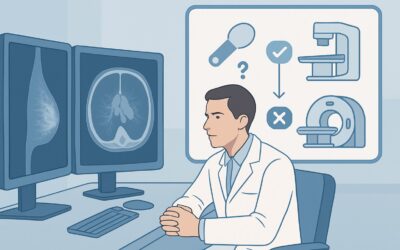
However, a bone scan alone is insufficient. The ACR also rates CT chest abdomen pelvis with IV contrast and FDG-PET/CT skull base to mid-thigh as Usually Appropriate. In practice, a bone scan is often paired with a contrast-enhanced CT to provide a comprehensive assessment of both the skeleton and the visceral organs.
Let’s compare the primary options:
- Bone Scan + CT CAP: This combination is a widely accepted standard. The bone scan excels at skeletal evaluation, while the CT provides detailed assessment of the lungs, liver, adrenal glands, and distant lymph nodes. The bone scan has a moderate radiation dose (☢☢☢ 1-10 mSv), while the CT carries a higher dose (☢☢☢☢ 10-30 mSv) and requires IV contrast.
- FDG-PET/CT: This single study evaluates the entire body for metabolically active disease in both bone and soft tissues. It can be more sensitive than conventional imaging for certain lesions, particularly lytic bone metastases. However, it may be less sensitive for indolent, purely osteoblastic metastases with low glucose uptake. Given its higher radiation dose (☢☢☢☢ 10-30 mSv) and cost, its use as a first-line tool varies by institution, often being reserved for more aggressive tumor subtypes or for clarifying equivocal findings from conventional imaging.
Modalities like MRI breast without and with IV contrast are rated Usually Not Appropriate for this indication because their purpose is to evaluate locoregional, not distant, disease. An MRI of the breast does not assess the skeleton, lungs, or liver.
Once you’ve decided on a Whole Body Bone Scan, our protocol guide covers the technique, radiotracer, and reading principles: Nuclear Medicine Bone Scan (Whole Body).
What’s Next After Staging Scans? Downstream Clinical Workflow
The results of the staging workup directly inform the patient’s treatment pathway. The downstream decisions diverge based on whether distant disease is identified.
If Staging Scans Are Positive for Metastases
If either the bone scan or CT scan shows findings classic for metastatic disease (e.g., multiple new osseous lesions, new liver masses), the patient is diagnosed with Stage IV breast cancer. The treatment plan shifts from curative-intent locoregional therapy (surgery and radiation) to systemic therapy (e.g., endocrine therapy plus a CDK4/6 inhibitor). If only a single, isolated lesion is found (oligometastatic disease), a biopsy may be performed to confirm metastasis, as this can have profound treatment implications.
If Staging Scans Are Negative
If both the bone scan and CT are negative for distant disease, the patient’s cancer is confirmed as clinical Stage IIB or III. They can proceed with a curative-intent treatment plan, which may include neoadjuvant (pre-operative) systemic therapy followed by surgery and radiation, or surgery first followed by adjuvant (post-operative) therapy.
If Staging Scans Are Indeterminate
Equivocal findings are common, such as a single “hot spot” on a bone scan that could represent arthritis or a small, nonspecific liver lesion on CT. In these cases, further investigation is required. This may involve targeted imaging (e.g., MRI of the specific bone) or proceeding to a problem-solving study like an FDG-PET/CT, which can often clarify if an indeterminate lesion is metabolically active and likely malignant. A multidisciplinary tumor board discussion is invaluable for resolving these complex cases.
Pitfalls to Avoid (and When to Get Help)
Navigating the staging process for late-stage breast cancer requires careful interpretation to avoid common errors.
- Over-calling Benign Findings: A bone scan is highly sensitive but not specific. Degenerative joint disease, healing fractures, and arthritis are frequent causes of increased radiotracer uptake. A radiologist’s interpretation, considering the pattern and location of findings, is crucial to avoid misdiagnosing benign changes as metastases.
- Incomplete Visceral Assessment: Relying solely on a bone scan provides an incomplete picture. For Stage IIB-III disease, the risk of visceral metastases is significant, making a contrast-enhanced CT of the chest, abdomen, and pelvis a necessary component of the workup.
- Misinterpreting the “Flare” Response: After initiating systemic therapy, healing bone metastases can paradoxically show increased uptake on a follow-up bone scan. This “flare phenomenon” can be mistaken for disease progression. Correlating with clinical symptoms and other imaging is essential.
If staging results are conflicting, ambiguous, or unexpected, the best course of action is to present the case at a multidisciplinary tumor board. This collaborative review by oncologists, radiologists, surgeons, and pathologists ensures a consensus on the patient’s definitive stage and optimal treatment plan.
Related ACR Topics and Tools
This article covers one specific workflow within the broader topic of breast cancer imaging. For a comprehensive overview of all clinical variants, from early-stage disease to surveillance, please consult our parent guide. You can also use the tools below to explore adjacent scenarios, review imaging protocols, and discuss radiation dose with your patients.
- For breadth across all scenarios in Imaging of Invasive Breast Cancer, see our parent guide: Imaging of Invasive Breast Cancer: ACR Appropriateness Decoded.
- ACR Appropriateness Criteria Lookup — for adjacent scenarios
- Imaging Protocol Library — for technique on the recommended study
- Radiation Dose Calculator — for cumulative dose conversations
Frequently Asked Questions
Why not just order an FDG-PET/CT for every patient with Stage IIB-III breast cancer?
While FDG-PET/CT is a powerful, single-session whole-body imaging tool, the combination of a bone scan and a contrast-enhanced CT of the chest, abdomen, and pelvis is also highly accurate for staging ER-positive breast cancer and is often more accessible. The ACR rates all three options as ‘Usually Appropriate,’ allowing for flexibility based on institutional preference, availability, and specific clinical factors. PET/CT is often reserved for clarifying equivocal findings from conventional imaging or for more aggressive subtypes like triple-negative or inflammatory breast cancer.
My patient has renal insufficiency. Can I still order a CT for visceral staging?
Yes, but the protocol must be modified. A non-contrast CT of the chest is sufficient for evaluating the lungs. For the abdomen and pelvis, a non-contrast CT can still detect many liver metastases and assess lymph nodes, though it is less sensitive than a contrast-enhanced study. An alternative for the abdomen is an MRI, potentially with a non-nephrotoxic gadolinium-based contrast agent. The decision requires a risk-benefit discussion, balancing diagnostic needs against the risks of contrast media.
The bone scan showed a single suspicious lesion. What is the next step?
A solitary bone lesion requires further investigation to avoid incorrectly upstaging a patient to Stage IV. The next step is typically targeted imaging with a modality that provides superior anatomical detail, such as a dedicated CT or MRI of the specific area. If imaging remains indeterminate, a percutaneous bone biopsy may be necessary to obtain a definitive histopathologic diagnosis.
Are these staging recommendations different for Invasive Lobular Carcinoma (ILC)?
No, for the purposes of distant disease staging, the recommendations are the same for both IDC and ILC. While ILC can be more subtle on mammography and has a propensity for unusual metastatic sites, it shares the high frequency of bone metastasis seen in ER-positive IDC. Therefore, a bone scan and cross-sectional imaging remain the standard of care for staging late-stage ILC.
Do these staging guidelines apply after a patient has completed neoadjuvant chemotherapy?
This workflow is specifically for initial staging at the time of diagnosis, before any treatment has been administered. Imaging performed after neoadjuvant therapy is for restaging and assessing treatment response, which is a different clinical question. The choice of imaging for restaging may differ and often involves the modality that best depicted the disease at baseline, which could include PET/CT or MRI.
Reviewed by Pouyan Golshani, MD, Interventional Radiologist — May 29, 2026