Which Imaging Study Best Evaluates Delayed Postoperative Dysphagia? ACR Guidelines

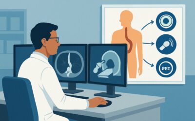
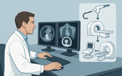
A 68-year-old woman is in your clinic, four months after an anterior cervical discectomy and fusion (ACDF). For the past few weeks, she’s felt that solid foods, particularly bread and meat, are “getting stuck” in her lower neck. Her initial postoperative recovery was uneventful, but this new symptom is causing significant anxiety. You suspect a delayed complication from her surgery, but the differential is broad. Which imaging study is the most appropriate first step to diagnose the cause of her dysphagia? This article provides a clinical workflow for this specific scenario, guided by the American College of Radiology (ACR) Appropriateness Criteria, which rates a Fluoroscopy single contrast esophagram as Usually Appropriate for this presentation.
Who Fits This Clinical Scenario?
This guidance applies to patients presenting with new or worsening dysphagia—either oropharyngeal (trouble initiating a swallow) or retrosternal (sensation of food sticking)—that develops more than one month after a surgical procedure. The prior surgery could involve the neck (like ACDF or thyroidectomy), thorax (like esophagectomy or cardiac surgery), or even the upper abdomen, as long as it could plausibly affect esophageal structure or function.
This scenario is distinct from several similar presentations that require a different diagnostic approach:
- Early Postoperative Dysphagia (less than 1 month): Dysphagia occurring immediately or within the first few weeks of surgery is often due to acute edema, hematoma, or nerve palsy. The workup and management for this early postoperative scenario may differ, focusing on more acute complications.
- Unexplained Dysphagia (No Surgical History): For a patient with no relevant surgical history, the pretest probability shifts toward primary esophageal disorders like achalasia, eosinophilic esophagitis, or an undiagnosed malignancy, altering the initial imaging choice.
- Dysphagia with a Known Cause: If the patient has a clear, attributable cause for their symptoms (e.g., a known Zenker’s diverticulum or a previously diagnosed stricture), the imaging may be for procedural planning rather than initial diagnosis.
This article focuses exclusively on the initial imaging workup for the delayed onset of symptoms in a postoperative patient, where the differential is heavily influenced by potential surgical complications.
What Diagnoses Are You Working Up in This Scenario?
In a patient with delayed postoperative dysphagia, the differential diagnosis centers on structural and functional changes related to healing, scarring, or hardware. The goal of initial imaging is to differentiate among these possibilities.
Benign Esophageal Stricture
This is a common cause of delayed postoperative dysphagia. Scar tissue formation at an anastomotic site (after an esophagectomy) or from localized inflammation and fibrosis related to surgery (e.g., from retraction during neck surgery) can cause gradual luminal narrowing. Symptoms typically progress from solids to liquids.
Recurrent Malignancy
If the index surgery was for esophageal, head and neck, or lung cancer, recurrence is a primary concern. Tumor regrowth can cause dysphagia through direct luminal invasion, extrinsic compression, or encasement of the esophagus, leading to a fixed narrowing or dysmotility.
Extrinsic Compression
Delayed complications like a chronic, organized hematoma, seroma, or abscess can compress the esophagus. More commonly, surgical hardware, such as an anterior cervical spine plate, can lead to chronic inflammation, fibrosis, or, in rare cases, erosion into the esophagus, causing a fistula.
Esophageal Motility Disorder
Injury to the vagus nerve or recurrent laryngeal nerve during neck or thoracic surgery can lead to disordered esophageal peristalsis or upper esophageal sphincter dysfunction. While less common to present in a delayed fashion, it remains a key consideration if structural imaging is normal.
Why Is Fluoroscopy Single Contrast Esophagram the Recommended Study?
The ACR rates both Fluoroscopy single contrast esophagram and CT of the neck and chest with IV contrast as Usually Appropriate for this scenario. However, the esophagram is often the preferred starting point because it directly visualizes luminal pathology and function in real-time, which are central to the most common differential diagnoses.
A single contrast esophagram provides excellent mucosal detail, making it highly sensitive for identifying benign strictures, webs, or the irregular narrowing characteristic of a mucosal malignancy. The dynamic nature of fluoroscopy allows for assessment of esophageal motility, revealing aperistalsis or tertiary contractions that suggest a functional disorder. It is the best initial test for mapping the length and severity of a stricture or identifying a potential fistula, which appears as a track of contrast extending outside the esophageal lumen.
While CT is also highly rated, its strengths lie elsewhere. CT neck and chest with IV contrast is superior for evaluating extra-esophageal pathology. It can precisely delineate an abscess, identify recurrent tumor in adjacent lymph nodes, or assess the relationship of surgical hardware to the esophagus. The choice between these two studies often depends on the leading clinical suspicion. If a luminal issue like a stricture is most likely, the esophagram is the logical first step. If extrinsic compression or a deep infection is suspected, CT may be preferred.
Why are other studies rated lower for this specific scenario?
- Fluoroscopy barium swallow modified (MBS): Rated May be appropriate. The MBS is a specialized study focused on the oropharyngeal phase of swallowing and aspiration risk. While useful if symptoms point specifically to the throat, it does not evaluate the entire thoracic esophagus, where many postoperative issues like strictures or fistulas occur.
- CT neck and chest without IV contrast: Rated Usually not appropriate. Omitting intravenous contrast severely limits the ability to assess for abscesses, enhancing tumors, or vascular complications. It provides anatomical information but misses the key soft-tissue and vascular detail needed to evaluate most of the serious differential diagnoses.
From a safety perspective, the esophagram involves a lower radiation dose (adult relative radiation level ☢☢☢, 1-10 mSv) compared to a chest CT (☢☢☢☢, 10-30 mSv), making it a favorable initial choice when clinically appropriate.
What’s Next After a Fluoroscopy Single Contrast Esophagram? Downstream Workflow
The results of the esophagram will guide your next steps in a branching clinical pathway. The goal is to move from diagnosis to management efficiently.
If the study is positive for a stricture:
A finding of luminal narrowing, consistent with a benign stricture or suspected malignancy, warrants a referral to a gastroenterologist. The next step is typically an esophagogastroduodenoscopy (EGD). EGD allows for direct visualization, biopsy to rule out malignancy, and therapeutic intervention, such as balloon or bougie dilation of a benign stricture.
If the study is positive for extrinsic compression or a suspected fistula:
When the esophagram suggests an external process is impinging on the esophagus or shows a tract of contrast leaving the lumen, the next step is a CT of the neck and chest with IV contrast. The CT will provide the detailed cross-sectional anatomy needed to characterize the compressing mass (e.g., fluid collection, tumor, hardware) or define the course of a fistula, which is critical for surgical or endoscopic planning.
If the study is negative:
A normal esophagram is reassuring but does not rule out all pathology. If the patient’s dysphagia persists, especially if it is mild or intermittent, the differential shifts. Consider an EGD to look for subtle mucosal abnormalities like eosinophilic esophagitis that may not be visible on fluoroscopy. If both the esophagram and EGD are normal, an esophageal manometry study may be indicated to formally diagnose a primary motility disorder.
Pitfalls to Avoid (and When to Get Help)
Navigating the workup for delayed postoperative dysphagia requires careful consideration to avoid common diagnostic errors.
- Incomplete Clinical History: Failing to provide the radiologist with the specific type of prior surgery, the date it was performed, and the nature of the patient’s symptoms (solids vs. liquids, location of sticking) can significantly limit the diagnostic utility of the esophagram.
- Attribution Error: While the prior surgery is the most likely culprit, it’s crucial to remember that the dysphagia could be from a new, unrelated condition. Keep a broad differential, especially if the initial imaging is unrevealing.
- Stopping the Workup Prematurely: A negative esophagram in a persistently symptomatic patient is not the end of the investigation. It effectively rules out significant structural blockages but should prompt a pivot to endoscopic evaluation or motility testing.
If a patient presents with “red flag” symptoms such as fever, chest pain, odynophagia (painful swallowing), or signs of sepsis, this suggests a more acute and serious complication like a contained perforation or abscess. In this case, you should escalate care immediately, often involving an urgent CT scan and consultation with GI and/or surgical services.
Related ACR Topics and Tools
For a comprehensive overview of imaging for all types of dysphagia, please see our parent guide. For tools to help you select the right study and understand the details, the following resources are available.
- For breadth across all scenarios in Dysphagia, see our parent guide: Dysphagia: ACR Appropriateness Decoded.
- To look up appropriateness criteria for adjacent or alternative clinical scenarios, use the ACR Appropriateness Criteria Lookup.
- For technical details on how specific studies are performed, consult the Imaging Protocol Library.
- To discuss cumulative radiation exposure with your patients, the Radiation Dose Calculator can be a helpful aid.
Frequently Asked Questions
Why is a single contrast esophagram preferred over a biphasic (double contrast) esophagram in this scenario?
A biphasic esophagram, which uses both thick barium and an effervescent agent to distend and coat the esophagus, is excellent for detecting subtle mucosal detail like early esophagitis or small cancers. However, in the delayed postoperative setting, the primary questions are about larger structural issues like strictures, fistulas, or extrinsic compression, for which a single contrast study is sufficient, simpler to perform, and often better tolerated by patients with significant dysphagia.
If my patient had neck surgery and now has a hoarse voice along with dysphagia, does that change the imaging plan?
Yes, the presence of hoarseness is highly suggestive of recurrent laryngeal nerve injury, a known complication of neck and thoracic surgery. While a fluoroscopic esophagram is still an appropriate first step to evaluate the swallowing mechanics and rule out a structural blockage, this finding increases the suspicion for a motility disorder. If the esophagram is normal, you may proceed more quickly to laryngoscopy and/or esophageal manometry.
Is there a role for MRI in evaluating delayed postoperative dysphagia?
Magnetic Resonance Imaging (MRI) is generally not a first-line imaging modality for dysphagia. Its primary strengths are in soft tissue characterization, particularly in the brain and musculoskeletal system. For the esophagus, fluoroscopy and CT provide better evaluation of the lumen, motility, and surrounding structures. MRI may have a niche role in complex cases, such as evaluating spinal cord or nerve root compression after spine surgery, but it is not part of the standard initial workup for the dysphagia itself.
My patient cannot tolerate oral contrast for the esophagram. What is the next best test?
If a patient cannot safely swallow contrast for a fluoroscopic study, the best alternative is a CT of the neck and chest with IV contrast. While it won’t provide the same functional information as an esophagram, it is excellent for identifying the most serious structural causes, such as extrinsic compression from a fluid collection, recurrent tumor, or hardware-related complications. An EGD would also be a reasonable next step, though it is more invasive.
Should I order the CT with oral contrast as well as IV contrast?
In this specific scenario, adding oral contrast to the CT scan can sometimes be helpful to better delineate the esophageal lumen and identify a fistula. However, it can also create streak artifact that obscures surrounding structures. The decision is often best left to the discretion of the radiologist. When ordering, it is most important to specify IV contrast and provide a detailed clinical history, allowing the imaging team to tailor the protocol for the highest diagnostic yield.
Reviewed by Pouyan Golshani, MD, Interventional Radiologist — May 29, 2026