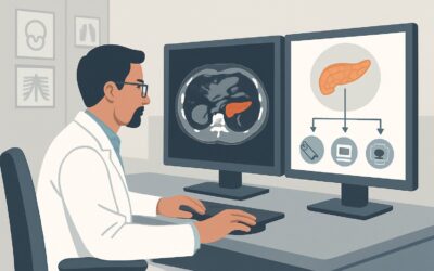
Which Imaging Study Is Best for a Palpable Mass Suspected to be an Intra-Abdominal Neoplasm?

A 62-year-old male with a history of chronic hepatitis C presents to your primary care clinic with a three-week history of vague abdominal fullness and a 10-pound weight loss. On examination, you palpate a firm, non-tender mass in the right upper quadrant. Your clinical suspicion is high for an intra-abdominal neoplasm, possibly related to his underlying liver disease. The immediate question is which imaging study to order first to confirm the presence of a mass, identify its organ of origin, and guide the subsequent workup. This article provides a focused clinical workflow for this exact scenario, based on the American College of Radiology (ACR) Appropriateness Criteria. For the initial imaging of a suspected intra-abdominal neoplasm, an abdominal ultrasound is rated Usually Appropriate.
Who Fits This Clinical Scenario?
This guidance applies to adult patients presenting with a palpable abdominal mass discovered on physical examination where there is a clinical suspicion for an underlying intra-abdominal neoplasm. The key factor is that the mass is believed to originate from an organ or structure within the peritoneal or retroperitoneal space, such as the liver, kidney, pancreas, or mesenteric lymph nodes.
This workflow is appropriate for the initial, undifferentiated workup. It assumes no prior relevant imaging is available.
It is crucial to distinguish this scenario from a similar but distinct clinical presentation:
- Suspected Abdominal Wall Mass: If the physical exam suggests the mass is superficial and moves with the contraction of the abdominal wall muscles (e.g., a positive Carnett’s sign), the mass is likely within the abdominal wall itself (e.g., rectus sheath hematoma, desmoid tumor, or soft tissue sarcoma). This represents a different clinical question and routes to the sibling ACR variant, “Palpable abdominal mass. Suspected abdominal wall mass. Initial imaging.”
- Known Primary Malignancy: If the patient has a known cancer and the palpable mass is suspected to be a metastasis, the imaging choice is typically guided by staging protocols for that specific cancer, which may favor PET/CT or contrast-enhanced CT from the outset.
- Acute Abdominal Pain: If the palpable mass is accompanied by acute pain, fever, or signs of peritonitis, the workup shifts toward an emergent evaluation for conditions like abscess, appendicitis, or diverticulitis, where CT is often the primary modality.
What Diagnoses Are You Working Up in This Scenario?
When a palpable intra-abdominal mass is suspected to be neoplastic, the differential diagnosis is broad and often guided by the mass’s location and the patient’s risk factors. The initial imaging aims to narrow this list by identifying the organ of origin and characterizing the lesion.
A primary consideration, especially for a right upper quadrant mass in a patient with liver disease, is a primary liver malignancy like hepatocellular carcinoma (HCC). Metastatic disease to the liver from an unknown primary (e.g., colorectal, pancreatic, or lung cancer) is also very common and can present as a palpable, enlarged liver or a discrete mass.
Another common origin is the kidney. A renal cell carcinoma (RCC) can grow quite large before becoming palpable, often presenting as a flank or upper quadrant mass. While many are found incidentally, a palpable mass can be the initial sign.
In the epigastrium, a pancreatic neoplasm, such as pancreatic ductal adenocarcinoma, can become palpable, particularly if it involves the body or tail of the pancreas or causes significant secondary effects like biliary obstruction. Similarly, lymphoma can present as bulky retroperitoneal or mesenteric lymphadenopathy that coalesces into a palpable mass.
Less common but important considerations include gastrointestinal stromal tumors (GISTs), primary sarcomas of the retroperitoneum, or neoplasms of the adrenal gland or spleen. Imaging also serves to identify non-neoplastic mimics, such as a large aortic aneurysm, splenomegaly from portal hypertension, or a simple benign cyst.
Why Is US abdomen the Recommended Initial Study for This Presentation?
The ACR designates both Ultrasound (US) abdomen and CT abdomen with IV contrast as Usually Appropriate for this scenario. However, ultrasound is often the superior initial examination due to its combination of safety, accessibility, and diagnostic utility for the primary clinical questions.
Rationale for Ultrasound (US) abdomen: Ultrasound is an excellent first-line tool for evaluating a palpable abdominal mass. Its primary strengths in this context are:
- Safety: It uses no ionizing radiation (0 mSv) and does not require IV contrast, avoiding potential risks of nephrotoxicity or allergic reaction.
- Characterization: It can rapidly differentiate a simple, benign cyst from a complex or solid mass that requires further investigation. This is often the most critical first step.
- Localization: US can typically identify the organ of origin (e.g., liver, kidney, gallbladder), which is fundamental to narrowing the differential diagnosis and planning the next steps.
- Accessibility and Cost: Ultrasound is widely available, relatively inexpensive, and can often be performed more quickly than CT or MRI.
Comparison to Alternatives:
- CT abdomen with IV contrast: While also rated Usually Appropriate, CT is generally reserved as the next step after an ultrasound confirms a solid or indeterminate mass. It provides superior global anatomic detail for staging and evaluating vascular involvement. However, it exposes the patient to ionizing radiation (☢☢☢ 1-10 mSv) and the risks of IV contrast. Using US first allows for a more targeted and justified use of CT.
- Radiography abdomen: This study is rated Usually Not Appropriate. Plain radiographs have very poor soft-tissue contrast and will fail to visualize most intra-abdominal neoplasms. They are only useful if they show secondary signs like organ displacement, abnormal gas patterns, or specific calcifications (e.g., in a mucinous adenocarcinoma), but the diagnostic yield is far too low to be a primary tool.
For these reasons, the logical workflow begins with the safest effective test. Ultrasound answers the key initial questions: Is there a mass? Is it cystic or solid? Where is it coming from? The answers then guide the use of more advanced, and higher-risk, imaging like CT or MRI.
What’s Next After US abdomen? Downstream Workflow
The results of the initial abdominal ultrasound will dictate the subsequent management and imaging pathway. The goal is to move from detection to characterization and, if necessary, staging.
- If the US is definitively positive for a solid mass: The next step is typically cross-sectional imaging tailored to the organ of origin.
- Solid Renal Mass: A dedicated multiphase CT or MRI of the abdomen is required to characterize the mass (e.g., Bosniak classification for cystic masses, characterization of solid enhancement for suspected RCC) and to stage for local invasion or metastatic disease. This is where a study like CT abdomen with IV contrast or MRI abdomen without and with IV contrast (May be appropriate) becomes essential.
- Suspicious Liver Lesion: A multiphase CT or MRI with liver-specific contrast agents is the standard of care for characterizing liver masses and is critical for diagnosis and staging of HCC or metastases.
- Other Solid Mass (e.g., Pancreatic, Retroperitoneal): Contrast-enhanced CT is generally the workhorse for further evaluation and staging.
- If the US is negative but clinical suspicion remains high: A negative ultrasound does not entirely rule out a neoplasm, especially for deep retroperitoneal structures or in patients with a large body habitus that limits sonographic visualization. In this case, proceeding directly to a CT abdomen with IV contrast is a reasonable and necessary next step to ensure a deep or obscured mass is not missed.
- If the US is indeterminate (e.g., complex cyst, ill-defined lesion): When ultrasound cannot confidently characterize a finding as benign, further imaging is required. MRI is often superior for problem-solving, particularly for liver lesions, complex renal cysts, and adrenal masses. Contrast-enhanced CT is also an excellent alternative.
Pitfalls to Avoid (and When to Get Help)
Navigating the workup of a palpable abdominal mass requires careful clinical correlation and an awareness of common diagnostic traps.
- Over-reliance on a “negative” ultrasound: In a patient with high-risk features (e.g., weight loss, anemia, specific risk factors) and a palpable mass, a negative or technically limited ultrasound should not end the workup. Have a low threshold to escalate to CT or MRI.
- Misattributing the organ of origin: A large left renal mass can be mistaken for splenomegaly, or a pancreatic tail mass can be mistaken for a primary adrenal or renal lesion. If the origin is unclear, cross-sectional imaging is mandatory.
- Ignoring body habitus: Ultrasound performance is significantly degraded by obesity and overlying bowel gas. If the sonographer reports a “technically limited study,” do not accept indeterminate findings; proceed to CT or MRI.
- Failing to order the correct contrast phase: Once you proceed to CT or MRI, ensure the protocol is appropriate for the suspected pathology. A generic “CT Abdomen/Pelvis with contrast” may not include the specific arterial, portal venous, and delayed phases needed to characterize a liver or renal mass.
If the imaging findings are complex or discordant with the clinical picture, consultation with the radiologist is invaluable. A direct conversation can help clarify the report and determine the most appropriate next imaging study or if biopsy is warranted.
Related ACR Topics and Tools
This article covers one specific scenario. For a comprehensive overview of all related presentations and for tools to help with ordering, see the resources below.
- For breadth across all scenarios in Palpable Abdominal Mass-Suspected Neoplasm, see our parent guide: Palpable Abdominal Mass-Suspected Neoplasm: ACR Appropriateness Decoded.
- ACR Appropriateness Criteria Lookup: Use this to explore evidence-based recommendations for thousands of clinical scenarios. ACR Appropriateness Criteria Lookup
- Imaging Protocol Library: Review detailed imaging techniques for the recommended studies. Imaging Protocol Library
- Radiation Dose Calculator: Discuss cumulative radiation exposure with patients when considering CT scans. Radiation Dose Calculator
Frequently Asked Questions
Why is ultrasound recommended over CT as the first step if CT is also rated ‘Usually Appropriate’?
Ultrasound is recommended first primarily due to its superior safety profile. It involves no ionizing radiation and typically requires no IV contrast, avoiding potential renal or allergic complications. It is highly effective for the initial goals: confirming a mass exists, differentiating cystic from solid, and identifying the organ of origin. CT is reserved as the next step for detailed characterization and staging once a solid or indeterminate mass is confirmed.
What if the patient is obese and I’m concerned about the quality of the ultrasound?
This is a critical clinical consideration. In patients with a large body habitus, ultrasound imaging can be technically limited, reducing its sensitivity. In such cases, it is reasonable to consider proceeding directly to CT abdomen with IV contrast as the initial imaging study, as it is less affected by body size and provides more definitive anatomical detail.
Does the location of the palpable mass (e.g., RUQ vs. epigastric) change the initial imaging choice?
No, for the initial evaluation, abdominal ultrasound remains the recommended first step regardless of the mass’s location within the abdomen. The location helps narrow the clinical differential diagnosis (e.g., RUQ suggests liver/gallbladder, epigastric suggests pancreas/stomach), but the imaging algorithm starts with the safest, most accessible modality capable of answering the primary questions.
If the ultrasound shows a simple cyst, is any further workup needed?
If an ultrasound confidently characterizes a finding as a simple benign cyst (e.g., in the liver or kidney) and this finding is consistent with the size and location of the palpable mass, no further imaging or workup for that finding is typically necessary. The palpable ‘mass’ was likely the benign cyst.
Should I order tumor markers like AFP or CA 19-9 before or after the initial imaging?
Tumor markers can be ordered concurrently with the initial imaging. They should not delay the imaging workup. The results of both the imaging and the lab tests are used together. For example, a solid liver mass on ultrasound in the setting of a very high AFP level is highly suggestive of hepatocellular carcinoma and would prompt an urgent, dedicated liver MRI or CT.
Reviewed by Pouyan Golshani, MD, Interventional Radiologist — May 29, 2026