Which Imaging Study Is Best for Late Postpartum Hemorrhage After Vaginal Delivery?

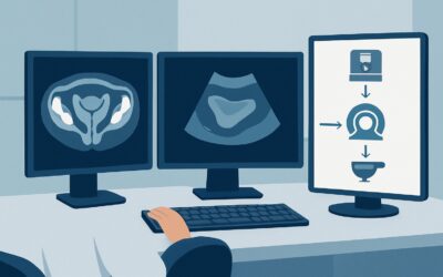
A 28-year-old woman presents to the emergency department with persistent, heavy vaginal bleeding three weeks after an uncomplicated vaginal delivery. She is hemodynamically stable but anxious about the ongoing blood loss. As the treating clinician, you suspect a cause beyond normal lochia, and your immediate question is which imaging study will most safely and effectively identify the source of this late postpartum hemorrhage. This article provides a detailed workflow for this specific clinical scenario, explaining why the American College of Radiology (ACR) Appropriateness Criteria panel rates `US duplex Doppler pelvis` as “Usually Appropriate” for the initial workup.
Who Fits This Clinical Scenario?
This guidance is for a specific patient population: women presenting with postpartum hemorrhage occurring in the late, or secondary, period. The inclusion criteria are precise:
- Timing: The hemorrhage occurs between 24 hours and 6 weeks after delivery.
- Delivery Type: The patient had a vaginal delivery.
- Presentation: This is the initial imaging workup for the bleeding episode.
It is crucial to distinguish this scenario from similar presentations that follow different diagnostic pathways. This workflow does not apply if:
- The hemorrhage is early (within the first 24 hours): Early postpartum hemorrhage after a vaginal delivery has a different differential, with uterine atony being the most common cause. The urgency and initial management steps differ significantly.
- The delivery was via cesarean section: Late postpartum hemorrhage after a cesarean delivery introduces surgical site complications to the differential, such as uterine dehiscence, hematoma, or abscess, which may alter the choice or interpretation of imaging.
Correctly identifying your patient within this specific context ensures the most appropriate and highest-yield imaging is ordered first.
What Diagnoses Are You Working Up in This Scenario?
When a patient presents with late postpartum hemorrhage after a vaginal delivery, imaging is directed at identifying a structural or vascular cause. The clinical differential diagnosis guides the choice of study and the radiologist’s interpretation.
Retained Products of Conception (RPOC) is the most common cause of secondary postpartum hemorrhage. This refers to placental or fetal tissue that remains in the uterus after delivery, acting as a nidus for bleeding and infection. On imaging, the goal is to identify an echogenic, intracavitary mass, often with demonstrable vascularity on Doppler imaging.
Subinvolution of the Placental Site is another primary consideration. This occurs when the uterus fails to contract and regress as expected after delivery. The placental implantation site can continue to bleed from exposed, non-constricted spiral arteries. Ultrasound can demonstrate a bulky, boggy uterus and help exclude other causes.
Endometritis, an infection of the uterine lining, can also cause late PPH. While primarily a clinical diagnosis based on fever, uterine tenderness, and foul-smelling lochia, imaging can reveal supportive findings like a thickened, heterogeneous endometrium, intrauterine fluid, or gas. More importantly, imaging helps rule out RPOC, which can coexist with or mimic infection.
Uterine Artery Pseudoaneurysm or Arteriovenous Malformation (AVM) are less common but are life-threatening causes of delayed hemorrhage. These vascular anomalies can result from vessel injury during delivery. They represent a critical diagnostic target because mismanagement (e.g., performing a D&C without a correct diagnosis) can lead to catastrophic bleeding. Doppler ultrasound is essential for their detection.
Why Is US duplex Doppler pelvis the Recommended Initial Study?
For a stable patient with late postpartum hemorrhage after a vaginal delivery, the ACR designates `US duplex Doppler pelvis`, `US pelvis transabdominal`, and `US pelvis transvaginal` as “Usually Appropriate.” The combination, particularly with Doppler, is the cornerstone of the initial workup for several compelling reasons.
First, ultrasound is highly sensitive for the most common etiologies. Transvaginal views provide high-resolution images of the endometrial cavity, ideal for identifying an echogenic mass concerning for Retained Products of Conception (RPOC). It can also assess for uterine subinvolution and signs of endometritis. Critically, the addition of color and spectral Doppler is not optional—it is essential. Doppler imaging can demonstrate a vascular stalk feeding a mass, significantly increasing the specificity for RPOC over a simple blood clot. Furthermore, Doppler is the primary modality for diagnosing dangerous vascular pathologies like uterine artery pseudoaneurysms or AVMs, which appear as a tangle of vessels with high-velocity, low-resistance flow.
Second, ultrasound involves no ionizing radiation (Radiation Level: O 0 mSv). This is a paramount safety consideration in a young, postpartum patient who may be breastfeeding. It is readily available, cost-effective, and can be performed quickly at the bedside if necessary.
In contrast, other modalities are rated lower for this initial presentation:
- CT Angiography (CTA) abdomen and pelvis with IV contrast is rated “May be appropriate.” It is reserved for hemodynamically unstable patients with suspected active arterial extravasation or when ultrasound is equivocal and a vascular abnormality is still suspected. Its primary role is often as a prelude to interventional radiology for embolization. The significant radiation dose (☢☢☢☢ 10-30 mSv) makes it unsuitable for a first-line test in a stable patient.
- MRI pelvis without and with IV contrast is rated “May be appropriate (Disagreement).” MRI offers superior soft tissue contrast and can be an excellent problem-solving tool, particularly in differentiating RPOC from avascular clot when ultrasound is indeterminate. However, it is more expensive, less accessible, and takes longer to perform, making it a secondary option rather than the initial imaging test of choice.
What’s Next After US duplex Doppler pelvis? Downstream Workflow
The results of the pelvic ultrasound will guide your next clinical steps. The downstream workflow depends on whether the findings are positive, negative, or indeterminate.
If the study is positive for Retained Products of Conception (RPOC): A definitive finding of a vascularized, echogenic mass in the endometrial cavity typically warrants gynecologic consultation for surgical management. The standard treatment is a suction dilation and curettage (D&C), often performed under hysteroscopic guidance to ensure complete removal and minimize uterine trauma.
If the study is positive for a uterine artery pseudoaneurysm or AVM: This is a radiologic emergency. The next step is an immediate consultation with Interventional Radiology. Attempting a D&C in this setting can lead to massive, life-threatening hemorrhage. The definitive treatment is typically transcatheter uterine artery embolization (UAE), which is highly effective at controlling the bleeding while preserving the uterus.
If the study is negative: A normal ultrasound in a patient with persistent bleeding suggests other causes, such as coagulopathy or subinvolution that is not apparent on imaging. The patient may be managed expectantly with uterotonic agents (e.g., methylergonovine) or hormonal therapy. If bleeding continues despite a negative ultrasound and conservative management, further investigation with hysteroscopy or MRI may be considered.
If the study is indeterminate: Sometimes, ultrasound cannot definitively distinguish between retained products and a simple blood clot. In these cases, clinical correlation is key. If suspicion for RPOC remains high, proceeding to hysteroscopy or ordering an MRI for better tissue characterization may be appropriate.
Pitfalls to Avoid (and When to Get Help)
Navigating the workup for late postpartum hemorrhage requires careful attention to detail to avoid common missteps.
- Pitfall 1: Ordering a pelvic US without Doppler. A grayscale-only ultrasound can easily miss a pseudoaneurysm or AVM and has lower specificity for RPOC. Always explicitly request “duplex Doppler” evaluation of the uterus.
- Pitfall 2: Inadequate clinical history. Failing to provide the date and type of delivery, estimated blood loss, and vital signs on the imaging requisition can hamper radiologic interpretation. Context is critical.
- Pitfall 3: Delaying resuscitation for imaging. In a hemodynamically unstable patient, the priority is always stabilization with fluids, blood products, and uterotonics. Imaging should not delay life-saving measures.
If the patient is hemodynamically unstable, has signs of a serious vascular injury on ultrasound, or if bleeding is refractory to initial management, escalate care immediately by involving obstetrics, interventional radiology, and critical care teams.
Related ACR Topics and Tools
This article focuses on a single clinical scenario. For a comprehensive overview of imaging for all presentations of postpartum hemorrhage and to explore related clinical questions, several resources are available.
- For breadth across all scenarios in Postpartum Hemorrhage, see our parent guide: Postpartum Hemorrhage: ACR Appropriateness Decoded.
- To explore other clinical variants and their corresponding ACR recommendations, use the ACR Appropriateness Criteria Lookup.
- For detailed technical specifications on how to perform the recommended study, consult the Imaging Protocol Library.
- To discuss radiation exposure with patients when considering CT, the Radiation Dose Calculator can help frame the conversation.
Frequently Asked Questions
Why is Doppler so important for a late postpartum hemorrhage ultrasound?
Color and spectral Doppler are critical because they assess blood flow. This allows the radiologist to distinguish between a simple, avascular blood clot and true retained products of conception (RPOC), which often have a vascular stalk. Most importantly, Doppler is the primary method for identifying dangerous but rare causes like a uterine artery pseudoaneurysm or an arteriovenous malformation (AVM), which require urgent intervention and contraindicate procedures like a D&C.
What if the patient is hemodynamically unstable? Should I still start with ultrasound?
In a hemodynamically unstable patient, the first priority is resuscitation (IV access, fluids, blood products). While a rapid bedside ultrasound can be useful to look for obvious causes, the patient may need to bypass other imaging and go directly to the operating room or interventional radiology suite. In some cases, a CT Angiography (CTA) may be performed to rapidly identify the source of bleeding before embolization, but this should not delay stabilization.
Can an ultrasound reliably differentiate retained products of conception from a blood clot?
It can be challenging. The presence of a distinct, echogenic mass with internal vascularity on Doppler is highly specific for RPOC. However, an avascular mass is indeterminate and could be either a clot or non-viable retained tissue. In these equivocal cases, clinical correlation, serial monitoring, or a problem-solving MRI may be necessary.
Is there any role for a plain radiograph (X-ray) in this scenario?
No. A plain radiograph of the pelvis has no role in the initial workup of late postpartum hemorrhage. It does not provide any useful information about the uterus, endometrium, or vascular structures and would only result in unnecessary radiation exposure and a delay in diagnosis.
If the ultrasound is negative but the patient continues to bleed, what is the next step?
If the initial high-quality transvaginal and Doppler ultrasound is negative and the patient remains stable, the focus shifts to non-structural causes. This includes medical management with uterotonics or hormonal therapy and checking for coagulopathies. If bleeding persists and is significant, further evaluation with hysteroscopy (to directly visualize the endometrial cavity) or MRI (to look for subtle RPOC or other myometrial abnormalities) may be considered in consultation with a gynecologist.
Reviewed by Pouyan Golshani, MD, Interventional Radiologist — May 29, 2026