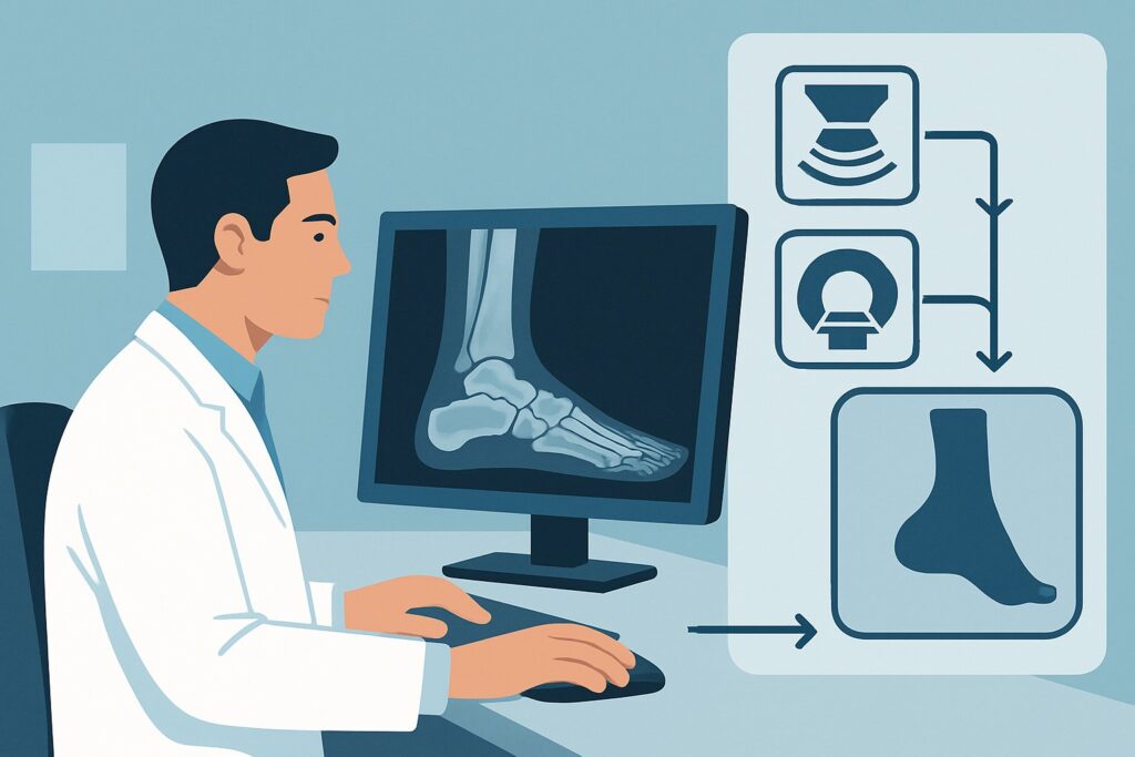
Which Imaging Study Is Best for Suspected Neurogenic Chronic Foot Pain?
A 48-year-old patient presents with six months of burning pain along the medial aspect of their heel and arch, worsening after long periods of standing. They describe a sensation of “pins and needles” that sometimes radiates toward the toes. Physical exam reveals tenderness over the proximal abductor hallucis muscle, but no obvious swelling or deformity. Plain radiographs of the foot were obtained two months ago and were interpreted as negative for fracture, arthritis, or other osseous abnormality. You suspect a neurogenic cause, like Baxter neuropathy or another nerve entrapment syndrome. What is the most appropriate next step in the imaging workup? This article provides a detailed clinical workflow for this specific scenario, grounded in the American College of Radiology (ACR) Appropriateness Criteria, which rates MRI of the foot without IV contrast as Usually Appropriate.
Who Fits This Clinical Scenario?
This guidance applies to adult patients with chronic foot pain (lasting three months or longer) where the clinical suspicion points toward a neurogenic origin. The key features of the presentation include symptoms like burning, tingling (paresthesia), numbness, or radiating pain that does not follow a clear musculoskeletal pattern. Crucially, this workflow is for patients who have already had initial radiographs that were either negative or indeterminate, effectively ruling out obvious osseous causes like stress fractures, significant degenerative disease, or bone tumors.
This scenario is distinct from other common causes of chronic foot pain. It is essential to differentiate it from presentations where:
- A soft-tissue injury is primary: If the pain is localized to a specific tendon, ligament, or the plantar fascia (e.g., classic plantar fasciitis symptoms without neuropathic features), the workup follows a different path.
- An occult fracture is suspected: If there is a history of trauma or repetitive stress with focal bony tenderness, the primary concern is an occult osseous injury, which has its own dedicated imaging algorithm.
- An osteochondral lesion is the lead diagnosis: If the pain is clearly articular, associated with locking or catching, and localized to a specific joint like the ankle or talus, the focus shifts to cartilage and subchondral bone.
This article is specifically for the patient whose history and exam suggest the pain generator is a nerve, whether from entrapment, inflammation, or a systemic process like complex regional pain syndrome (CRPS).
What Diagnoses Are You Working Up in This Scenario?
When ordering imaging for suspected neurogenic foot pain, you are primarily investigating conditions that cause nerve irritation, compression, or abnormal signaling. The differential diagnosis guides the choice of imaging modality and what the radiologist will be looking for.
Baxter Neuropathy (Inferior Calcaneal Nerve Entrapment): This is a frequent and often underdiagnosed cause of chronic heel pain, accounting for a significant portion of cases that fail to respond to treatment for plantar fasciitis. The inferior calcaneal nerve, the first branch of the lateral plantar nerve, can become compressed as it passes between the abductor hallucis and quadratus plantae muscles. Imaging can reveal signs of denervation, such as edema or fatty atrophy, in the abductor digiti minimi muscle, which this nerve innervates.
Tarsal Tunnel Syndrome: This condition involves the compression of the posterior tibial nerve or its branches as they pass through the tarsal tunnel, a fibro-osseous canal on the medial side of the ankle. Causes can include ganglion cysts, varicosities, fibrous bands, or post-traumatic changes. The symptoms are often broader than Baxter neuropathy, involving the heel, arch, and toes.
Complex Regional Pain Syndrome (CRPS): CRPS is a challenging diagnosis characterized by disproportionate pain, sensory changes, and autonomic dysfunction, often following a minor injury. While a clinical diagnosis, imaging can provide supportive evidence. The findings can be diffuse and non-specific but may include patchy bone marrow edema, soft tissue edema, and periarticular osteopenia, which are well-visualized on MRI.
Other Nerve Entrapments: Less commonly, other nerves such as the sural nerve (lateral foot) or superficial peroneal nerve (dorsal foot) can become entrapped. Imaging helps identify space-occupying lesions or post-traumatic fibrosis that may be responsible for the compression.
Why Is MRI of the Foot Without IV Contrast the Recommended Study?
The ACR designates MRI of the foot without intravenous contrast as Usually Appropriate for this clinical scenario because of its superior soft-tissue resolution and ability to directly visualize nerve pathology and its secondary effects without the use of ionizing radiation.
MRI provides a detailed anatomical map of the foot and ankle. It can directly identify potential causes of nerve compression, such as space-occupying lesions (e.g., ganglion cysts, schwannomas) or post-traumatic scarring. More importantly, it can detect the downstream consequences of nerve entrapment. In the case of Baxter neuropathy, the key finding is often edema or, in chronic cases, fatty atrophy of the abductor digiti minimi muscle. This sign of denervation is highly specific and can confirm the diagnosis when clinical suspicion is high.
For CRPS, MRI is sensitive for detecting the characteristic patchy or diffuse bone marrow edema and soft-tissue edema that support the diagnosis. This is particularly valuable when the clinical picture is ambiguous. The lack of ionizing radiation (Adult RRL: 0 mSv) is a significant advantage for a non-emergent workup in an adult patient.
Alternative studies are rated lower for specific reasons:
- Ultrasound (US) of the foot is rated May be appropriate. High-resolution ultrasound can visualize nerves and surrounding structures, and dynamic imaging can sometimes demonstrate nerve subluxation. However, it is highly operator-dependent, has a limited field of view, and is less sensitive than MRI for detecting bone marrow edema (a key feature of CRPS) or subtle muscle denervation changes.
- A 3-phase bone scan is also rated May be appropriate. It is sensitive for the increased vascularity and bone turnover seen in CRPS, often showing diffuse periarticular uptake in the delayed phase. However, the findings are non-specific, and the study involves a moderate radiation dose (Adult RRL: ☢☢☢ 1-10 mSv) without providing the detailed anatomical information of an MRI.
- CT of the foot, with or without contrast, is rated Usually not appropriate. While excellent for bone detail, it provides poor soft-tissue contrast and cannot adequately visualize nerves or early muscle denervation, making it unsuitable for this indication.
Initially, intravenous contrast is not typically required for this workup, as the key findings of nerve compression (muscle atrophy/edema) and CRPS (bone marrow edema) are well-visualized on non-contrast sequences. This avoids the risks and costs associated with gadolinium-based contrast agents.
What’s Next After MRI? Downstream Workflow
The results of the MRI will guide the subsequent management plan. The workflow branches based on whether the findings are positive, negative, or indeterminate.
If the MRI is positive for nerve entrapment: A definitive finding, such as denervation atrophy of the abductor digiti minimi muscle confirming Baxter neuropathy, solidifies the diagnosis. The next steps typically involve conservative management, including physical therapy, orthotics, and activity modification. If these fail, an image-guided anesthetic and corticosteroid injection may be considered for both diagnostic confirmation and therapeutic relief. The ACR rates this procedure as May be appropriate (Disagreement), reflecting variability in practice. For refractory cases, surgical nerve decompression may be the final option.
If the MRI is negative: A normal MRI in the face of persistent, high-suspicion clinical symptoms does not entirely rule out a neurogenic cause. The next step is often to pursue electrodiagnostic testing (nerve conduction studies and electromyography) to assess nerve function directly. A negative imaging study also prompts a re-evaluation of the differential diagnosis. Could the pain be from an early tendinopathy not yet visible on MRI, or a small-fiber neuropathy that has no imaging correlate? The clinical context becomes paramount.
If the MRI is indeterminate: Findings like mild, non-specific soft tissue or muscle edema require careful clinical correlation. These can be seen with overuse and may not be related to a primary nerve pathology. In this situation, a period of conservative management followed by a repeat clinical evaluation is often the most prudent course. If CRPS is suspected but MRI findings are equivocal, a 3-phase bone scan could be considered as a complementary study.
Pitfalls to Avoid (and When to Get Help)
Navigating the workup for neurogenic foot pain requires avoiding several common pitfalls. First, avoid anchoring on a diagnosis of plantar fasciitis for all heel pain; consider Baxter neuropathy in patients who fail standard treatments. Second, do not order a CT scan for this indication, as it lacks the necessary soft-tissue resolution and exposes the patient to unnecessary radiation. Third, remember that a negative MRI does not mean the patient’s pain is not real; it may simply mean the pathology is functional (requiring electrodiagnostics) rather than structural. Finally, be precise in your order, specifying the suspected diagnosis (e.g., “rule out Baxter neuropathy”) to help the radiologist tailor the imaging protocol and focus their interpretation. If the diagnosis remains elusive after initial imaging and conservative care, referral to a physiatrist, neurologist, or foot and ankle specialist is the appropriate next step for further evaluation and management.
Related ACR Topics and Tools
This article focuses on a single, specific clinical scenario. For a comprehensive overview of all variants within this topic, please consult our parent guide. The tools below can help you apply evidence-based guidelines to other clinical questions.
- For breadth across all scenarios in Chronic Foot Pain, see our parent guide: Chronic Foot Pain: ACR Appropriateness Decoded.
- ACR Appropriateness Criteria Lookup — for adjacent scenarios
- Imaging Protocol Library — for technique on the recommended study
- Radiation Dose Calculator — for cumulative dose conversations
Frequently Asked Questions
Why is MRI without contrast preferred over MRI with contrast for suspected Baxter neuropathy?
For suspected nerve entrapment syndromes like Baxter neuropathy, the key diagnostic findings are often secondary signs of muscle denervation—specifically, edema in the acute phase and fatty atrophy in the chronic phase within the abductor digiti minimi muscle. These changes are well-visualized on standard non-contrast T1-weighted and fluid-sensitive sequences. Intravenous contrast is typically not needed to see these findings and is reserved for cases where a mass, tumor, or abscess is suspected as the cause of compression.
If I strongly suspect Complex Regional Pain Syndrome (CRPS), is a 3-phase bone scan a better first choice than MRI?
While a 3-phase bone scan is sensitive for CRPS, the American College of Radiology (ACR) still lists MRI as ‘Usually Appropriate’ and the bone scan as ‘May be appropriate’ for this scenario. MRI provides superior anatomical detail, can identify alternative diagnoses that may mimic CRPS, and avoids ionizing radiation. A bone scan can be a useful problem-solving tool if the MRI is equivocal or if there is a contraindication to MRI, but MRI is generally the preferred initial advanced imaging study.
What if the patient has a pacemaker or other contraindication to MRI?
If a patient cannot undergo an MRI, the next best steps depend on the leading diagnosis. High-resolution ultrasound is rated ‘May be appropriate’ and can be an excellent alternative for evaluating specific nerve entrapment sites, like the tarsal tunnel or the course of the inferior calcaneal nerve. If CRPS is the primary concern, a 3-phase bone scan would be the most appropriate alternative. The choice should be guided by the specific clinical question.
Can ultrasound be used to guide an injection for Baxter neuropathy?
Yes. Ultrasound is an excellent modality for guiding diagnostic and therapeutic injections around nerves. The ACR rates image-guided injections as ‘May be appropriate (Disagreement)’ for this scenario. If an injection is planned, ultrasound allows for real-time visualization of the needle tip to ensure accurate placement of anesthetic and/or corticosteroid around the inferior calcaneal nerve, improving efficacy and safety.
My patient’s symptoms sound like neurogenic pain, but they also have tenderness directly over the plantar fascia insertion. How does that change the workup?
This is a common clinical challenge, as Baxter neuropathy and plantar fasciitis can coexist and have overlapping symptoms. If radiographs are negative, an MRI without contrast remains the best single test to evaluate both conditions. It can clearly show thickening and signal abnormality of the plantar fascia while simultaneously assessing for muscle denervation changes that would indicate a nerve entrapment. This allows for a comprehensive evaluation from one study.
Reviewed by Pouyan Golshani, MD, Interventional Radiologist — May 30, 2026