Which Imaging Study Is Best for Suspected Recurrent Melanoma? ACR Workflow

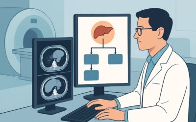
A 58-year-old patient with a history of a Clark level IV melanoma excised from his left upper back two years ago presents to your clinic. During a self-exam, he noticed a new, firm, non-tender lump in his left axilla. On your examination, you palpate a 1.5 cm mobile nodule consistent with a lymph node. You are concerned about regional recurrence, but reactive lymphadenopathy is also possible. The immediate clinical question is which imaging study to order first to evaluate this finding and guide the next steps in staging. For this specific presentation, the American College of Radiology (ACR) Appropriateness Criteria designates Ultrasound (US) of the area of interest as a Usually appropriate initial imaging test.
Who Fits This Clinical Scenario for Suspected Melanoma Recurrence?
This guidance is for a specific and common clinical situation: an adult patient with a previously diagnosed and treated cutaneous, muco-cutaneous, or ocular melanoma who now presents with new signs or symptoms concerning for recurrence. The key element is a new clinical finding—such as a palpable lymph node, a new subcutaneous nodule near the old scar, or symptoms suggesting distant spread—that triggers a workup for suspected regional or metastatic disease. This is fundamentally a restaging evaluation.
This workflow is distinct from other melanoma imaging scenarios. It does not apply to:
- Initial Staging: A patient with a newly diagnosed primary melanoma who has no clinical signs or symptoms of regional or metastatic disease. That situation follows a different diagnostic algorithm based on the primary tumor’s characteristics.
- Asymptomatic Surveillance: A patient with a history of melanoma who is undergoing routine, scheduled follow-up imaging without any new, concerning physical exam findings or symptoms.
- Initial Staging with Known High-Risk Features: A patient with a new melanoma diagnosis who already has microscopic satellites identified on the initial biopsy specimen. While related, that scenario has its own specific imaging considerations from the outset.
Applying this guidance correctly means focusing on patients where a new clinical suspicion of recurrence has emerged after a disease-free interval.
What Diagnoses Are You Working Up in This Scenario?
When a patient with a history of melanoma develops a new localized finding, the differential diagnosis is focused but includes both malignant and benign possibilities. The goal of imaging is to characterize the finding and determine the need for tissue sampling.
Regional Nodal Metastasis: This is the most pressing concern. Melanoma has a high propensity for lymphatic spread, and the regional nodal basin (e.g., axilla, groin, neck) is the most common first site of recurrence. A new, firm, or enlarging lymph node in a draining basin for the primary tumor site is highly suspicious for a metastasis until proven otherwise.
In-Transit or Satellite Metastasis: These represent tumor cells that have spread from the primary site via lymphatics but have not yet reached a regional lymph node. Satellite metastases are found within 2 cm of the primary tumor scar, while in-transit metastases are located more than 2 cm away but before the next draining nodal basin. They typically present as new cutaneous or subcutaneous nodules.
Benign Reactive Lymphadenopathy: Lymph nodes can enlarge for many reasons unrelated to cancer, including local infection, skin inflammation, or systemic viral illness. While a less likely explanation in a high-risk patient, it remains a key differential. Imaging can help distinguish reactive features (preserved fatty hilum, oval shape) from malignant ones.
Second Primary Malignancy: Less commonly, a new nodule could represent an entirely different disease process. This could include a second primary cancer like lymphoma, which manifests as lymphadenopathy, or a metastasis from another, undiagnosed primary tumor. While not the leading diagnosis, it is a possibility if the biopsy results are inconsistent with melanoma.
Why Is Ultrasound of the Area of Interest the Recommended First Study?
For evaluating a specific, palpable area of concern in a patient with a history of melanoma, ultrasound is the ideal initial imaging modality. The ACR rates ‘US area of interest’ as Usually appropriate because it directly answers the immediate clinical question with high accuracy and efficiency, while also facilitating the definitive next step.
The primary strength of ultrasound in this context is its excellent spatial resolution for superficial soft tissues. It can precisely characterize a palpable nodule, confirming if it is a lymph node, a subcutaneous deposit, or another type of lesion. For lymph nodes, sonographic features such as a rounded shape, loss of the normal fatty hilum, eccentric cortical thickening, and increased vascularity are highly suggestive of metastatic involvement. This morphologic detail is often superior to that provided by CT for superficial nodes.
Most importantly, ultrasound is a real-time imaging tool that enables immediate image-guided biopsy. If a suspicious node or nodule is identified, a fine-needle aspiration (FNA) or core needle biopsy can be performed during the same appointment. This ability to move directly from characterization to tissue sampling is a significant workflow advantage, providing a rapid, definitive diagnosis that guides all subsequent systemic staging and treatment decisions. Furthermore, US involves no ionizing radiation (RRL=O 0 mSv) and does not require IV contrast, making it safe and widely accessible.
In contrast, other powerful imaging studies are rated lower as the first step for this specific problem:
- FDG-PET/CT whole body is also rated Usually appropriate for systemic staging, but it is not the best initial test for a focal, palpable finding. It involves significant radiation (RRL=☢☢☢☢ 10-30 mSv) and is less effective at guiding a biopsy of a specific superficial nodule than US. PET/CT is typically reserved for comprehensive systemic staging after a recurrence has been pathologically confirmed via a US-guided biopsy.
- Radiography chest is rated Usually not appropriate. Its sensitivity for detecting early pulmonary metastases is very low, and it provides no information about the soft tissues or nodal basins where recurrence is suspected. A CT of the chest would be the appropriate study if lung metastases are a specific concern.
When ordering, be specific: “Ultrasound of the [e.g., left axilla] to evaluate palpable nodule. Please assess lymph node morphology and be prepared for possible US-guided FNA/biopsy.”
What’s Next After Ultrasound? Downstream Workflow
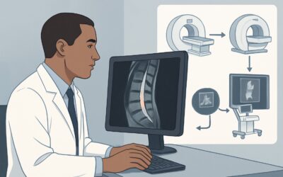
The results of the ultrasound and any associated biopsy will dictate the subsequent management pathway. The workflow branches based on whether the findings are positive, negative, or indeterminate for recurrent melanoma.
If the US-guided biopsy is POSITIVE for melanoma: This confirms recurrent disease. The patient now requires full systemic restaging to determine the overall extent of disease, as this will guide treatment decisions (e.g., surgery, immunotherapy, targeted therapy). This is the point at which a more comprehensive study, such as FDG-PET/CT whole body, becomes Usually appropriate. An MRI of the brain without and with IV contrast is also often performed, as melanoma has a high propensity for central nervous system metastases.
If the US is negative or shows benign features (and biopsy is negative/not performed): If the ultrasound demonstrates clearly benign features, such as a reactive-appearing lymph node with a preserved fatty hilum, and clinical suspicion is low, a short-term clinical or imaging follow-up may be sufficient. If a biopsy is performed and is negative for malignancy, this is reassuring. However, if clinical suspicion remains high despite a negative FNA, a core or excisional biopsy may still be warranted to rule out a false negative.
If the US is indeterminate and biopsy is non-diagnostic: Sometimes, imaging features are equivocal, and an FNA sample may be insufficient for a definitive diagnosis. In this situation, the next step is typically to obtain more tissue. This usually involves a US-guided core needle biopsy or a surgical excisional biopsy of the suspicious lymph node or nodule. Proceeding to systemic imaging like PET/CT is generally not indicated until a tissue diagnosis is secured.
Pitfalls to Avoid (and When to Get Help)
Navigating the workup for suspected melanoma recurrence requires careful attention to detail to avoid common missteps.
- Pitfall 1: Skipping the Biopsy. Do not proceed to full-body systemic staging based on suspicious imaging features alone. Pathologic confirmation of recurrence is critical before initiating major therapeutic interventions. Ultrasound’s key advantage is enabling that biopsy.
- Pitfall 2: Ordering the Wrong Initial Study. Jumping directly to a PET/CT for a single palpable nodule is often an inefficient use of resources. It exposes the patient to unnecessary radiation and is less effective than US for guiding a biopsy of a superficial lesion. Start with the targeted, problem-solving study first.
- Pitfall 3: Accepting a Non-Diagnostic FNA. If a fine-needle aspiration is inconclusive but clinical and sonographic suspicion for recurrence remains high, do not stop the workup. The next step should be a core needle biopsy or excisional biopsy to obtain a definitive histologic diagnosis.
If you encounter a complex case with discordant clinical, imaging, and pathologic findings, this is the time to escalate. A discussion with the reporting radiologist and a referral to a multidisciplinary tumor board with surgical oncology, medical oncology, and pathology is the appropriate next step.
Related ACR Topics and Tools
For a comprehensive overview of imaging across all clinical variants of melanoma staging and follow-up, as well as tools to help with study selection and patient communication, the following resources are available.
- For breadth across all scenarios in Staging and Follow-up of Melanoma, see our parent guide: Staging and Follow-up of Melanoma: ACR Appropriateness Decoded.
- To explore other clinical presentations and their corresponding ACR recommendations, use the ACR Appropriateness Criteria Lookup.
- For detailed procedural techniques on recommended studies, consult the Imaging Protocol Library.
- To discuss radiation exposure from studies like CT and PET/CT with your patients, the Radiation Dose Calculator can help quantify cumulative dose.
Frequently Asked Questions
Why not order a PET/CT scan first for a suspected melanoma recurrence?
While PET/CT is excellent for whole-body staging, it’s not the best initial test for a focal, palpable finding. Ultrasound is more sensitive for evaluating superficial lymph node morphology, involves no radiation, and, most importantly, allows for immediate image-guided biopsy to confirm the diagnosis. The standard workflow is to confirm recurrence with a US-guided biopsy first, then use PET/CT for systemic staging.
What if the patient has multiple palpable nodules in different areas?
If there are multiple areas of concern (e.g., nodules in the axilla and near the primary scar), ultrasound is still the appropriate first step. The radiologist can evaluate all clinically relevant sites in one session and potentially biopsy the most accessible or suspicious lesion. If findings suggest widespread disease, this further strengthens the case for subsequent systemic staging with PET/CT after confirmation.
Is an MRI an acceptable alternative to ultrasound for a palpable nodule?
MRI is not typically used as the first-line study for a palpable superficial nodule. While it provides excellent soft tissue contrast, it is more expensive, less accessible, and does not offer the real-time biopsy guidance that ultrasound does. MRI is rated ‘Usually appropriate’ for specific indications like evaluating the abdomen/pelvis or brain for metastases, but not for the initial workup of a palpable superficial lesion.
What if the suspected recurrence is for ocular melanoma?
This guidance applies to suspected regional or distant recurrence from any type of melanoma, including ocular. If an ocular melanoma survivor develops a suspicious nodule in the neck or a palpable liver lesion, for example, the same principles apply. The initial study should be targeted to the clinical finding—often an ultrasound of the neck or abdomen—to obtain a tissue diagnosis before proceeding to full systemic staging.
If the US-guided biopsy is negative, is melanoma recurrence ruled out?
A negative biopsy is highly reassuring, but not 100% definitive, especially with a fine-needle aspiration (FNA) which can have a false-negative rate. If the clinical and sonographic suspicion for malignancy remains high despite a negative FNA, the next step should be a core needle biopsy or a surgical excisional biopsy to obtain more tissue and rule out sampling error.
Reviewed by Pouyan Golshani, MD, Interventional Radiologist — May 30, 2026