Which Imaging Study Is Best for Traumatic Hip Pain with Suspected Soft Tissue Injury?
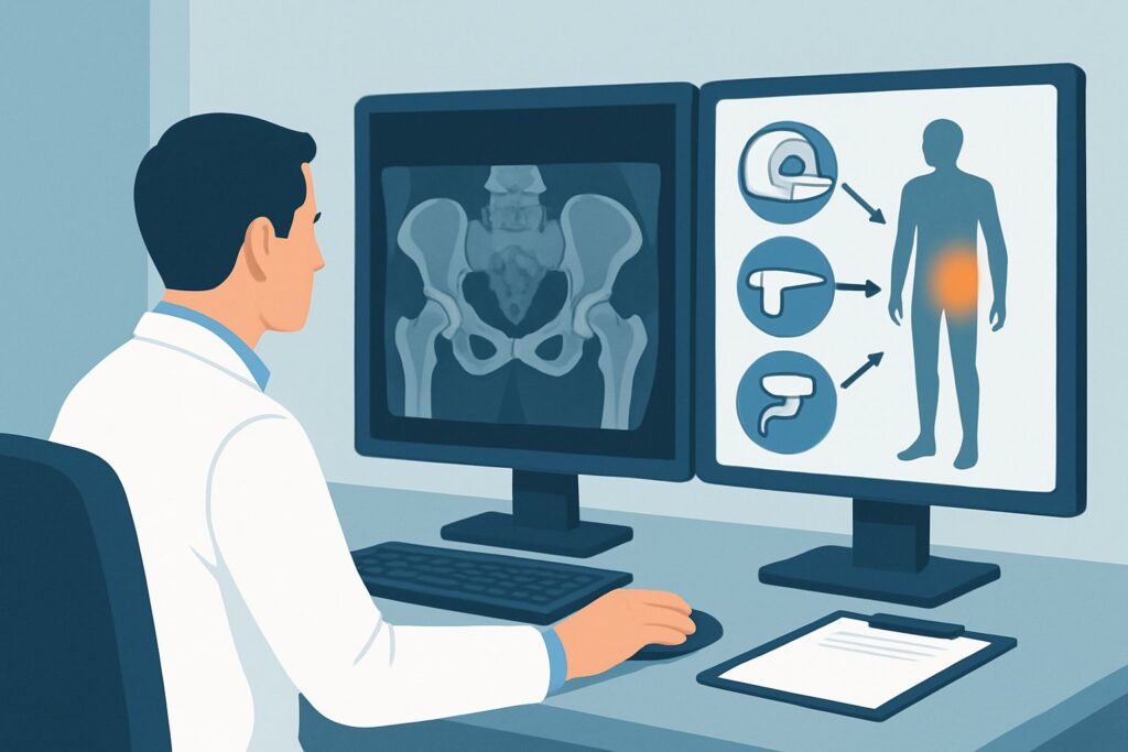
A 38-year-old patient presents to your clinic with acute left hip pain after a weekend soccer match. They describe a sudden, sharp pain during a sprint, and now they are limping with significant discomfort over the lateral hip and buttock. You obtained radiographs in the emergency department, which were read as negative for fracture or dislocation. Your clinical suspicion is high for a significant soft tissue injury, such as a gluteal tendon tear or a proximal hamstring avulsion. The immediate question is how to confirm the diagnosis and guide treatment. This article provides a step-by-step workflow for this specific scenario, grounded in the American College of Radiology (ACR) Appropriateness Criteria. For an adult with traumatic hip pain, negative radiographs, and suspected tendon, muscle, or ligament injury, an MRI hip without IV contrast is rated Usually Appropriate.
Who Fits This Clinical Scenario for Traumatic Hip Pain?
This guidance is tailored for a precise clinical situation. It applies to adult patients who meet all the following criteria:
- Acute, Traumatic Onset: The hip pain began suddenly due to a specific event, such as a fall, sports injury, or forceful movement.
- Negative or Indeterminate Radiographs: Initial X-rays have already been performed and do not show a clear fracture, dislocation, or other osseous cause for the pain.
- Suspicion for Soft Tissue Injury: The clinical examination and history point toward an injury of the muscles, tendons, or ligaments around the hip. This includes findings like focal tenderness away from bone, pain with specific muscle activation, or a palpable defect.
It is critical to distinguish this scenario from similar presentations that require a different diagnostic approach. This workflow does not apply if:
- You highly suspect an occult fracture: If the patient has significant risk factors (e.g., osteoporosis, high-energy trauma) and focal bony tenderness despite negative radiographs, the primary concern is a radiographically occult fracture. This follows a different ACR variant.
- This is the initial imaging study: If no imaging has been performed yet, radiographs are almost always the appropriate first step for acute traumatic hip pain.
- The pain is non-traumatic or chronic: This guidance is not intended for the workup of gradual-onset hip pain, osteoarthritis, or chronic tendinopathy.
What Diagnoses Are You Working Up in This Scenario?
When initial radiographs are negative, the focus shifts to the soft tissue structures that stabilize and move the hip. The differential diagnosis in this setting is broad, and the goal of advanced imaging is to pinpoint the exact location and severity of the injury.
Gluteal Tendon Tear: Often involving the gluteus medius or minimus tendons at their insertion on the greater trochanter, this is a very common cause of lateral hip pain, sometimes referred to as greater trochanteric pain syndrome. Acute traumatic tears can occur from falls or sudden twisting motions and are a key diagnosis to confirm or exclude.
Proximal Hamstring Tendon Injury: An acute tear or avulsion of the hamstring origin from the ischial tuberosity typically results from forceful hip flexion with the knee extended (e.g., sprinting, hurdling, or water skiing). Patients present with deep buttock pain and weakness with knee flexion.
Iliopsoas Strain or Tear: Injury to the primary hip flexor muscle-tendon unit causes pain in the anterior hip and groin. This can happen during kicking motions or other activities involving rapid, forceful hip flexion.
Acetabular Labral Tear: While often associated with chronic conditions like femoroacetabular impingement, the labrum can be torn acutely during trauma. Patients may report mechanical symptoms like clicking or locking in addition to sharp groin pain.
Ligamentous Injury (Hip Sprain): Significant trauma, such as a subluxation that spontaneously reduced, can cause sprains or tears of the hip capsule and its reinforcing ligaments (e.g., iliofemoral, pubofemoral, ischiofemoral ligaments) or the ligamentum teres. These are often injuries of exclusion but can be visualized on advanced imaging.
Why Is MRI Hip Without IV Contrast the Recommended Study for Suspected Soft Tissue Injury?
The ACR designates MRI hip without IV contrast as Usually Appropriate for this scenario because it directly addresses the clinical question with the highest diagnostic accuracy and no ionizing radiation.
The primary strength of Magnetic Resonance Imaging (MRI) is its superior soft tissue contrast. It can clearly delineate individual muscles, tendons, ligaments, and cartilage, making it the ideal modality to identify tears, strains, inflammation, and edema. Fluid-sensitive sequences (like T2-weighted or proton density-weighted images with fat suppression) are exceptionally good at highlighting the edema and fluid that accompany acute traumatic injuries, effectively acting as a natural contrast agent.
For this reason, intravenous contrast is typically unnecessary. The information needed to diagnose an acute muscle or tendon tear is readily available on a non-contrast study. Adding IV contrast increases the cost, scan time, and introduces the rare risks associated with gadolinium-based agents without providing significant additional diagnostic value for this specific indication.
Why are other studies rated lower?
- US hip (Usually not appropriate): While ultrasound is a valuable tool for evaluating superficial structures and can be used for dynamic assessment, it has significant limitations in this context. It is highly operator-dependent, provides a limited field of view, and struggles to visualize deep structures of the hip, such as the hamstring origin or the acetabular labrum. It cannot provide the comprehensive anatomical survey needed after trauma.
- CT hip (Usually not appropriate): Computed Tomography (CT) is excellent for evaluating complex bone anatomy but offers poor visualization of soft tissues. It cannot reliably diagnose tendon, muscle, or labral tears. Furthermore, CT exposes the patient to ionizing radiation (adult_rrl=☢☢☢ 1-10 mSv), which is unwarranted when a non-radiation modality like MRI (adult_rrl=O 0 mSv) is diagnostically superior for the clinical question.
Once you’ve decided on MRI hip without IV contrast, our protocol guide covers the technique, contrast, and reading principles: MRI Hip Without Contrast.
What’s Next After the MRI? Downstream Workflow
The results of the MRI will directly guide your next steps in management, which can range from conservative care to surgical consultation.
- If the MRI is positive for a significant tear (e.g., full-thickness gluteus medius tear, retracted hamstring avulsion): This finding often warrants a referral to an orthopaedic surgeon. The severity and location of the tear, along with the patient’s age and activity level, will determine whether surgical repair is indicated. The MRI provides the surgeon with the essential anatomical information needed for operative planning.
- If the MRI is positive for a low-grade injury (e.g., muscle strain, partial-thickness tendon tear, tendinosis): These injuries are typically managed non-operatively. The next step is a referral to physical therapy for a structured rehabilitation program. Pain management with activity modification and non-steroidal anti-inflammatory drugs (NSAIDs) is also a cornerstone of treatment.
- If the MRI is negative: A negative MRI is highly reassuring and effectively rules out a significant soft tissue injury. In this case, the diagnosis is likely a minor sprain or contusion that will resolve with rest and conservative care. If pain persists despite a negative MRI, you may need to reconsider the differential diagnosis to include non-structural causes of pain or referred pain from the lumbar spine.
- If the MRI is indeterminate or reveals an unexpected finding: Occasionally, the findings may be unclear or point toward a different pathology (e.g., stress fracture, avascular necrosis, or a tumor). In these cases, the next step is often a discussion with the interpreting radiologist to correlate the imaging with the clinical picture and determine if further imaging or a different specialty consultation is needed.
Pitfalls to Avoid (and When to Get Help)
Navigating this workup requires careful attention to the clinical details to avoid common missteps.
- Misattributing pain to “bursitis”: Before advanced imaging, lateral hip pain is often labeled as trochanteric bursitis. However, MRI studies have shown that underlying gluteal tendinopathy or tears are a far more common cause. Relying on a bursitis diagnosis without a proper workup can delay appropriate treatment.
- Ordering the wrong MRI: Be specific on your order. Requesting “MRI hip without contrast” is key. Adding contrast is unnecessary for this indication and increases cost.
- Ignoring the possibility of an occult fracture: If the mechanism was high-energy or the patient has osteoporosis, maintain a high index of suspicion for a fracture even with negative radiographs. If clinical suspicion for fracture is the primary driver, the imaging choice may be different (often MRI or CT, depending on the specific scenario).
- Delaying imaging for high-grade injuries: For injuries like a complete, retracted hamstring avulsion, early diagnosis and surgical repair can lead to better outcomes. A significant delay can make the repair more difficult due to tendon retraction and scarring.
If you identify a complete tendon avulsion, large full-thickness tear, or any unexpected aggressive-appearing lesion on MRI, escalate with an urgent referral to an orthopaedic surgeon.
Related ACR Topics and Tools
This article focuses on one specific clinical pathway. For a comprehensive overview of all scenarios, alternative imaging choices, and further resources, the following tools are invaluable.
For breadth across all scenarios in Acute Hip Pain, see our parent guide: Acute Hip Pain: ACR Appropriateness Decoded.
- ACR Appropriateness Criteria Lookup: Look up any clinical scenario to see the latest evidence-based imaging recommendations.
- Imaging Protocol Library: Access detailed technical protocols for performing the recommended imaging studies.
- Radiation Dose Calculator: Help patients understand radiation exposure from different medical imaging procedures.
Frequently Asked Questions
Why not just start with an MRI instead of radiographs for a traumatic hip injury?
Radiographs are the appropriate first step because they are fast, widely available, and excellent for identifying fractures and dislocations, which are the most urgent diagnoses to rule out after significant trauma. An MRI is a more resource-intensive study reserved for when radiographs are negative but a significant soft tissue or occult bone injury is still suspected.
Is an MR arthrogram better than a non-contrast MRI for this scenario?
No. An MR arthrogram involves injecting contrast directly into the hip joint and is primarily used to evaluate intra-articular structures, especially the acetabular labrum, in cases of chronic pain or instability. For an acute traumatic injury where extra-articular muscle and tendon tears are the main concern, a standard non-contrast MRI is sufficient and less invasive.
What if the patient has a contraindication to MRI, like a non-compatible pacemaker?
If MRI is contraindicated, the next best step is less clear and requires clinical judgment. A high-resolution ultrasound performed by an experienced musculoskeletal sonographer may be able to evaluate specific superficial tendons (like the gluteus medius). CT is poor for soft tissues but could be considered if an occult fracture is a competing concern. This situation often warrants a consultation with a radiologist to determine the best alternative.
How soon after the injury should the MRI be performed?
For most suspected low-grade to moderate soft tissue injuries, the MRI can be performed on a non-emergent outpatient basis within a few days to a week. However, if you suspect a high-grade, retractable injury (like a complete hamstring avulsion) that may require urgent surgical repair, the MRI should be performed more expeditiously to facilitate a prompt surgical consultation.
Does this guidance apply to adolescent patients with similar injuries?
While the principles are similar, adolescents have unique considerations, such as open growth plates (physes) and the risk of apophyseal avulsion fractures. These avulsion injuries can mimic soft tissue injuries clinically but are bony injuries. While MRI is still an excellent tool, the interpretation and differential diagnosis are different, and consultation with a pediatric radiologist or orthopedist is often beneficial.
Reviewed by Pouyan Golshani, MD, Interventional Radiologist — May 30, 2026