What Is the Best Next Imaging Study for a Hand Fracture with Suspected Soft Tissue Injury?
It’s a busy shift in the emergency department when you see a 24-year-old who punched a wall in frustration. The radiographs are back, confirming an oblique fracture of the fifth metacarpal neck—a classic boxer’s fracture. On physical exam, however, you notice subtle extensor lag at the metacarpophalangeal (MCP) joint and tenderness directly over the extensor hood. This isn’t just a simple fracture; you suspect a concomitant sagittal band or extensor tendon injury. The immediate clinical question is clear: what is the right next imaging study to evaluate the soft tissues without delaying care?
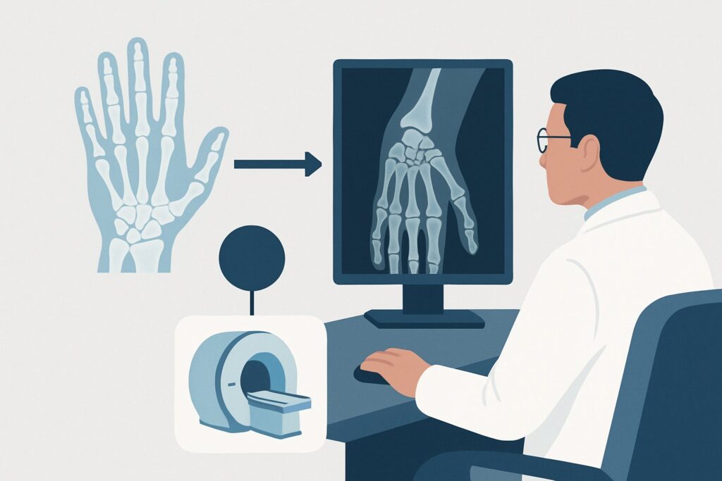
For this specific scenario—an acute hand fracture seen on radiographs with clinical suspicion for tendon or ligament trauma—the American College of Radiology (ACR) Appropriateness Criteria rate US hand as Usually Appropriate. This article provides a focused workflow for this presentation, detailing the rationale for this choice and the downstream clinical pathway.
Who Fits This Clinical Scenario for Suspected Hand Tendon or Ligament Injury?
This guidance is tailored for a very specific patient presentation. Correctly identifying if your patient fits this scenario is the crucial first step to ordering the most effective imaging.
Inclusion criteria for this workflow:
- The patient has experienced acute trauma to the hand.
- Initial radiographs have already been performed and confirm a fracture of a metacarpal or phalanx.
- The clinical examination suggests an associated soft tissue injury, such as a tendon tear, ligament rupture, or pulley injury. This suspicion may arise from findings like joint instability, tendon subluxation with motion, a palpable defect, or a specific mechanism of injury (e.g., hyperextension, rotational force).
This workflow does NOT apply if:
- Initial radiographs are negative or equivocal. If you suspect a fracture or soft tissue injury but the x-rays are clear, you are in a different clinical scenario that follows a separate diagnostic algorithm.
- The injury is primarily to the wrist. If a radiograph shows a distal radius or carpal bone fracture with suspected carpal ligament or triangular fibrocartilage complex (TFCC) injury, that constitutes a distinct scenario with different imaging recommendations.
- The primary finding is joint malalignment without a fracture. A subluxed or dislocated joint on radiographs in the absence of a fracture also follows a separate ACR-guided pathway.
Properly categorizing the patient presentation ensures that the chosen imaging study has the highest probability of answering the clinical question.
What Diagnoses Are You Working Up with Further Imaging?
When a hand fracture is accompanied by signs of soft tissue damage, the differential diagnosis expands beyond simple bone healing. The goal of subsequent imaging is to identify these consequential injuries that can significantly alter management, often requiring surgical intervention.
Extensor Tendon and Sagittal Band Injury
This is a common concern, especially with metacarpal fractures like the boxer’s fracture. The sagittal bands are crucial components of the extensor hood that centralize the extensor tendon over the MCP joint. A tear can cause the tendon to subluxate or dislocate, leading to weakness and dysfunction. Imaging is used to confirm the tear and assess the degree of tendon displacement.
Flexor Tendon and Pulley System Injury
While less common with closed fractures, flexor tendon injuries can occur. More frequently, the flexor pulley system, which holds the tendons close to the bone, can be damaged. An A2 or A4 pulley rupture, for example, can lead to tendon bowstringing and loss of mechanical advantage. Imaging helps visualize the integrity of these critical structures.
Collateral Ligament Rupture
Ligamentous injuries often accompany intra-articular or avulsion fractures. A classic example is an ulnar collateral ligament (UCL) tear at the thumb MCP joint (Skier’s or Gamekeeper’s thumb), which may be associated with an avulsion fracture at the base of the proximal phalanx. Identifying a complete tear, particularly with displacement (a Stener lesion), is critical as it is an indication for surgery.
Volar Plate Injury
Hyperextension injuries, especially at the proximal interphalangeal (PIP) joint, can cause a volar plate avulsion fracture. While the fracture is seen on radiographs, imaging can assess the degree of ligamentous retraction and associated collateral ligament damage, which informs the decision between conservative management and surgical repair.
Why Is Ultrasound of the Hand a Recommended Next Step?
The ACR rates both US hand and MRI hand without IV contrast as Usually Appropriate for this scenario. However, ultrasound often emerges as the more practical and diagnostically powerful first choice due to its unique capabilities.
The primary strength of ultrasound is its ability to perform dynamic imaging. A skilled sonographer can actively move the patient’s digit while scanning to visualize tendon subluxation, ligamentous stress-testing for laxity, or the formation of a gap in a torn tendon. This real-time functional information is invaluable for assessing injuries like sagittal band ruptures and cannot be replicated by static imaging modalities like MRI. Furthermore, ultrasound offers superb spatial resolution for the superficial structures of the hand, allowing for detailed evaluation of tendon fibers, ligaments, and pulleys. It involves no ionizing radiation (0 mSv) and allows for direct correlation of imaging findings with the patient’s point of maximal tenderness.
How do alternative studies compare?
- MRI hand without IV contrast is also Usually Appropriate and provides excellent anatomical detail of soft tissues, bone marrow, and occult fractures. It is a powerful tool, especially if ultrasound is equivocal or if a deeper or more complex injury pattern is suspected. However, it is a static examination, is typically more expensive, and may be less readily available than ultrasound.
- CT hand (with or without contrast) is rated Usually Not Appropriate. While CT excels at defining complex fracture patterns, its soft tissue contrast is poor, making it unsuitable for directly visualizing tendon or ligament tears. It also exposes the patient to a small amount of ionizing radiation (adult RRL ☢ <0.1 mSv).
- MRI hand with IV contrast is also Usually Not Appropriate. Gadolinium-based contrast is generally not required to assess for acute traumatic tendon or ligament injuries and adds unnecessary cost and potential risk.
- Bone scan is rated Usually Not Appropriate. This is a physiologic study that is highly sensitive for bone turnover but lacks the anatomic specificity needed to diagnose an acute tendon or ligament tear. It also carries a significant radiation dose (adult RRL ☢☢☢ 1-10 mSv).
For these reasons, ultrasound is often the most efficient and effective next step to characterize a suspected soft tissue injury in the setting of a known hand fracture.
What Is the Downstream Workflow After a Hand Ultrasound?
The results of the hand ultrasound will guide the next phase of management, which often involves collaboration with a hand surgeon. The post-imaging decision tree is relatively straightforward.
- If the ultrasound is positive for a significant injury: A definitive finding, such as a full-thickness tendon rupture, a displaced ligament tear (e.g., Stener lesion), or a functionally significant pulley rupture, typically warrants an urgent or semi-urgent consultation with a hand surgeon. The detailed information from the ultrasound report on the location and extent of the injury is crucial for surgical planning.
- If the ultrasound is negative: If the study shows no evidence of a significant tendon or ligament injury, the patient can often be managed conservatively for their fracture, with appropriate splinting and follow-up. The focus of care remains on fracture healing.
- If the ultrasound is equivocal or clinical suspicion remains high: In cases where the ultrasound is technically limited (e.g., by patient pain or swelling) or the results are indeterminate, but your clinical suspicion for a surgically significant injury is high, the next appropriate step is to obtain an MRI hand without IV contrast. MRI can serve as a powerful problem-solving tool, offering a comprehensive anatomical survey that may reveal a subtle tear or an alternative diagnosis missed on ultrasound.
This structured approach ensures that patients with significant soft tissue injuries are identified promptly for specialized care, while those without can proceed with standard fracture management.
Common Pitfalls to Avoid in This Hand Injury Scenario
Navigating this clinical scenario effectively requires avoiding a few common missteps that can lead to diagnostic delays or suboptimal outcomes.
- Pitfall 1: Defaulting to MRI first. Ordering a static MRI when the primary clinical question is one of dynamic instability (e.g., tendon subluxation) can miss the diagnosis. Ultrasound’s real-time capability is purpose-built for this question.
- Pitfall 2: Ordering CT for soft tissues. Using CT to evaluate a suspected tendon or ligament tear is a low-yield strategy. It exposes the patient to radiation without providing the necessary soft tissue detail. Reserve CT for cases where you need to better characterize a complex, comminuted, or intra-articular fracture pattern.
- Pitfall 3: Underestimating the clinical exam. A well-documented physical exam is key. Failing to test for tendon subluxation or joint instability may lead you to miss the indication for further soft tissue imaging altogether, treating only the fracture.
When to Escalate: If the patient presents with signs of an open fracture, compartment syndrome (tense compartment, pain with passive stretch, paresthesias), or acute neurovascular compromise, this constitutes a surgical emergency. Obtain immediate hand surgery consultation, as this supersedes any non-urgent imaging decisions.
Related ACR Topics and Tools
For further reading on related scenarios and to explore imaging decision support tools, the following resources are available. For breadth across all scenarios in Acute Hand and Wrist Trauma, see our parent guide: Acute Hand and Wrist Trauma: ACR Appropriateness Decoded.
- ACR Appropriateness Criteria Lookup — for adjacent scenarios
- Imaging Protocol Library — for technique on the recommended study
- Radiation Dose Calculator — for cumulative dose conversations
Frequently Asked Questions
Why is MRI also rated ‘Usually Appropriate’ if ultrasound is the top recommendation?
MRI is also rated ‘Usually Appropriate’ because it provides outstanding anatomical detail of soft tissues, bone, and bone marrow. It is an excellent alternative to ultrasound and is the preferred next step if an ultrasound is inconclusive or if a deeper, more complex injury is suspected. However, ultrasound is often favored first due to its lower cost, wider availability, and unique ability to perform dynamic, real-time assessment of tendon and ligament function, which is often the key clinical question.
What if the initial radiograph was negative but I still suspect a ligament injury?
That is a different clinical scenario (‘Suspect acute hand or wrist trauma. Initial radiographs negative or equivocal. Next imaging study.’). This article’s workflow applies only when a fracture has already been confirmed on radiographs. For patients with negative x-rays, the imaging workup is different and often proceeds directly to MRI to look for an occult fracture or primary soft tissue injury.
Does this guidance apply to chronic hand pain after a healed fracture?
No, this guidance is specifically for the acute trauma setting. Chronic pain after a fracture involves a different differential diagnosis, including nonunion, malunion, post-traumatic arthritis, or complex regional pain syndrome. The imaging workup for chronic pain follows a different pathway.
Should I order contrast with the MRI if I choose that path?
No. For this specific scenario, the ACR rates MRI *without* IV contrast as ‘Usually Appropriate’ and MRI *with* IV contrast as ‘Usually Not Appropriate.’ Intravenous contrast is not typically necessary to evaluate for acute traumatic tendon or ligament tears and adds potential risk and cost without providing significant additional diagnostic information.
What specific information should I provide the radiologist when ordering the hand ultrasound?
To maximize the diagnostic yield of the ultrasound, provide specific clinical details in your order. Include the precise location of tenderness, the specific injury you are trying to rule out (e.g., ‘evaluate for sagittal band rupture and extensor tendon subluxation at the 5th MCP joint’), the mechanism of injury, and the relevant findings from your physical exam. This context allows the sonographer and radiologist to tailor the dynamic exam to answer your clinical question.
Reviewed by Pouyan Golshani, MD, Interventional Radiologist — May 29, 2026