Which Procedure Is Best for Bleeding Gastric Varices with a Gastrorenal Shunt?

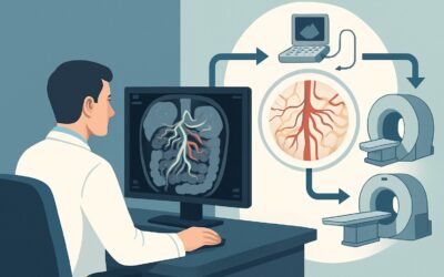
A 58-year-old male with known cirrhosis is admitted from the emergency department with hematemesis. Endoscopy confirms active bleeding from large gastric varices. His Model for End-stage Liver Disease (MELD) score is 12, and a recent MRI clearly delineates a large, high-flow gastrorenal shunt. A subsequent hepatic venous pressure gradient measurement is 10 mmHg. As the interventional radiologist on call, you must decide on the definitive management strategy. This is a common but critical decision point where choosing the right procedure can significantly impact both immediate hemostasis and long-term outcomes. This article details the clinical workflow for this specific presentation, where the underlying anatomy and relatively preserved liver function guide the therapeutic choice. For this patient, the American College of Radiology (ACR) Appropriateness Criteria rate Balloon-occluded Retrograde Transvenous Obliteration (BRTO) as a Usually Appropriate procedure.
Who Fits This Clinical Scenario?
This guidance applies to a well-defined patient cohort: individuals with liver cirrhosis presenting with bleeding from large, high-flow gastric varices. The key inclusion criteria that make this scenario distinct are the combination of a relatively low MELD score (around 12), a low-to-moderate degree of portal hypertension (indicated by a hepatic wedge pressure of 10 mmHg), and the confirmed presence of a large gastrorenal shunt on prior cross-sectional imaging like MRI or CT.
It is crucial to distinguish this presentation from similar but distinct clinical situations that require a different approach. This workflow is NOT intended for:
- Patients with very high MELD scores (e.g., >18-20): In a patient with a MELD of 20, the need for portal decompression is more urgent, and the risks of any procedure are higher. In that case, a Transjugular Intrahepatic Portosystemic Shunt (TIPS) might be more strongly considered despite the shunt anatomy.
- Patients with refractory hepatic encephalopathy: The presence of significant hepatic encephalopathy is a relative contraindication for TIPS. While BRTO does not typically worsen encephalopathy, the patient’s overall clinical status changes the risk-benefit calculation for all interventions.
- Patients without a demonstrable spontaneous portosystemic shunt: If no large gastrorenal or other shunt is identified, the technical approach for BRTO is not feasible. Management would then rely on endoscopic therapy or TIPS.
What Pathophysiology Are You Addressing in This Scenario?
In this patient, the primary diagnosis—bleeding from gastric varices—is already established. The clinical workup focuses on understanding the underlying hemodynamics driving the hemorrhage to select the most effective and least morbid treatment. The presence of a large gastrorenal shunt is the central finding that dictates the therapeutic algorithm.
Gastric varices are dilated submucosal veins that act as a collateral pathway for blood to bypass the high-pressure portal system and return to the systemic circulation. A gastrorenal shunt is a common and often large-caliber example of such a pathway, directing blood from the gastric veins (often via the left gastric or posterior gastric veins) into the left renal vein and subsequently the inferior vena cava. While this decompresses the portal system to a degree, the high flow and pressure through the thin-walled varices create a significant risk of life-threatening rupture.
The key insight in this scenario is that the bleeding is a direct consequence of this specific, well-defined anatomic conduit. The relatively low MELD score and hepatic wedge pressure suggest that the patient’s overall portal hypertension is not yet severe enough to mandate a global decompressive procedure like TIPS. The problem is localized to the shunt and the varices it feeds. Therefore, the therapeutic goal shifts from lowering overall portal pressure to directly obliterating the high-risk variceal complex by targeting its outflow shunt.
Why Is BRTO a Recommended Procedure for This Presentation?
For a cirrhotic patient with bleeding gastric varices, a low MELD score, modest portal hypertension, and a large gastrorenal shunt, Balloon-occluded Retrograde Transvenous Obliteration (BRTO) is rated as Usually Appropriate by the ACR. This procedure is uniquely suited to the pathophysiology of this specific scenario.
BRTO involves advancing a catheter, typically via a femoral or jugular vein approach, into the inferior vena cava, the left renal vein, and retrogradely into the gastrorenal shunt. A balloon is inflated to occlude the shunt’s outflow, and a sclerosant agent is injected to fill and thrombose the variceal complex. The balloon remains inflated for a period to allow the sclerosant to work without being washed out into the systemic circulation. This approach directly targets the bleeding source without creating a new intrahepatic shunt, thereby preserving hepatic perfusion and avoiding the risk of new or worsened hepatic encephalopathy. The high technical and clinical success rates for achieving hemostasis in patients with this anatomy make it a primary consideration.
Other procedures are also rated for this scenario, but BRTO often holds a clinical advantage here:
- TIPS: Also rated Usually Appropriate, TIPS creates an intrahepatic shunt to decompress the entire portal system. While effective at reducing variceal pressure, it is arguably “too much” procedure for a patient with a wedge pressure of only 10 mmHg. The primary indication for TIPS is managing the complications of severe portal hypertension, which this patient does not fully manifest. Furthermore, TIPS carries a notable risk of inducing or worsening hepatic encephalopathy, a complication that BRTO avoids.
- Endoscopic Management: Also Usually Appropriate, endoscopic therapy with agents like cyanoacrylate glue is often the first-line intervention to achieve initial hemostasis. However, for large, high-flow varices fed by a major shunt, the durability of endoscopic treatment can be limited, with higher rates of re-bleeding compared to definitive shunt obliteration with BRTO. It is often best viewed as a crucial bridge to a more definitive procedure.
BRTO is a fluoroscopically-guided procedure and involves ionizing radiation and iodinated contrast. The pre-procedural MRI or CT is essential for mapping the complex anatomy of the shunt and identifying any other significant draining veins that may need to be embolized for a successful outcome.
What’s Next After BRTO? Downstream Workflow
The post-procedure workflow is focused on confirming treatment success and monitoring for potential complications. The decision tree branches based on the immediate and short-term outcomes.
If BRTO is technically successful: The patient is typically monitored in a hospital setting for 24-48 hours. Key concerns include post-procedural pain, hematuria (from sclerosant irritation), and, more seriously, sclerosant migration or non-target embolization. A follow-up non-contrast CT scan is often performed before discharge or within the following week to confirm complete thrombosis of the gastric varices. The patient’s underlying liver disease and portal hypertension still require long-term management by a hepatologist, but the immediate bleeding threat from the treated varices is resolved. It is important to monitor for potential worsening of esophageal varices or ascites, as occluding a large decompressive shunt can transiently increase portal pressure.
If BRTO is not technically feasible: In some cases, the anatomy of the gastrorenal shunt may be too tortuous to navigate, or other outflow veins may prevent effective occlusion. If BRTO fails, the next logical step is to proceed with TIPS, which is also rated Usually Appropriate and provides an alternative, highly effective method for variceal decompression.
If bleeding recurs after BRTO: Recurrent bleeding is uncommon but can occur due to recanalization of the treated varices or the development of new varices from different feeding vessels. The workup would involve repeat endoscopy and cross-sectional imaging to understand the new anatomy, followed by consideration for repeat embolization or TIPS.
Pitfalls to Avoid (and When to Get Help)
Several potential pitfalls can complicate the management of this specific clinical scenario. First, failing to appreciate the significance of the low hepatic wedge pressure (10 mmHg) could lead to prematurely choosing TIPS, unnecessarily exposing a patient with relatively mild portal hypertension to the risk of hepatic encephalopathy. Second, inadequate pre-procedural imaging can lead to technical failure; a high-quality, multiphasic CT or MRI is critical to map all afferent and efferent vessels of the variceal system. Third, post-BRTO, it is crucial to monitor for worsening ascites or the development/enlargement of esophageal varices, as blocking a major portosystemic shunt can redirect portal flow. If a patient develops significant new-onset ascites or evidence of esophageal variceal bleeding post-BRTO, this represents a clinical escalation that requires urgent consultation with hepatology and consideration for TIPS.
Related ACR Topics and Tools
This article covers a single, specific variant within the broader topic of managing gastric varices. For a comprehensive overview of all clinical scenarios and their corresponding ACR ratings, please see our parent guide. Additional GigHz tools can help you navigate adjacent clinical questions and standardize your imaging requests.
- For breadth across all scenarios in Radiologic Management of Gastric Varices, see our parent guide: Radiologic Management of Gastric Varices: ACR Appropriateness Decoded.
- Imaging Appropriateness Selector — for adjacent scenarios
- Imaging Protocol Library — for technique on the recommended study
- Radiation Dose Calculator — for cumulative dose conversations
Frequently Asked Questions
Why is the MELD score of 12 so important in this scenario?
A MELD score of 12 indicates relatively preserved synthetic liver function. This makes the patient a better candidate for an intervention like BRTO and suggests that a major portal decompressive procedure like TIPS, with its associated risks of shunting blood away from the liver, is not immediately necessary.
What if the hepatic wedge pressure was 18 mmHg instead of 10 mmHg?
A higher hepatic wedge pressure (e.g., 18 mmHg) indicates more severe portal hypertension. In that case, even with a large gastrorenal shunt, the argument for a TIPS procedure becomes much stronger. TIPS would not only treat the gastric varices but also address the underlying high portal pressure, reducing the risk of other complications like ascites or esophageal variceal bleeding.
Can BRTO be performed if the patient has a splenorenal shunt instead of a gastrorenal shunt?
Yes, the BRTO technique can be adapted for other large portosystemic shunts, including splenorenal shunts. The principle remains the same: access the shunt retrogradely from the venous system, occlude the outflow with a balloon, and inject a sclerosant to thrombose the varices. The procedure is sometimes referred to as BATO (Balloon-occluded Antegrade Transvenous Obliteration) or CARTO (Coil-Assisted Retrograde Transvenous Obliteration) depending on the specific anatomy and technique used.
Does BRTO cure the patient’s portal hypertension?
No, BRTO is a targeted treatment for a specific complication of portal hypertension (bleeding gastric varices fed by a shunt). It does not treat the underlying liver disease or reduce the overall portal pressure. In fact, by closing a large decompressive shunt, it can transiently increase portal pressure, potentially worsening ascites or esophageal varices, which requires careful follow-up.
Is an MRI necessary if a high-quality contrast-enhanced CT has already been performed?
No, a high-quality, multiphasic contrast-enhanced CT is generally sufficient for pre-procedural planning for BRTO. Both modalities can effectively delineate the complex vascular anatomy of the portosystemic shunt and associated varices. The key is having one high-quality cross-sectional imaging study to serve as a roadmap for the intervention.
Reviewed by Pouyan Golshani, MD, Interventional Radiologist — May 26, 2026