Should You Order EBUS for a New 1-3 cm Hilar Lung Nodule in a Smoker?
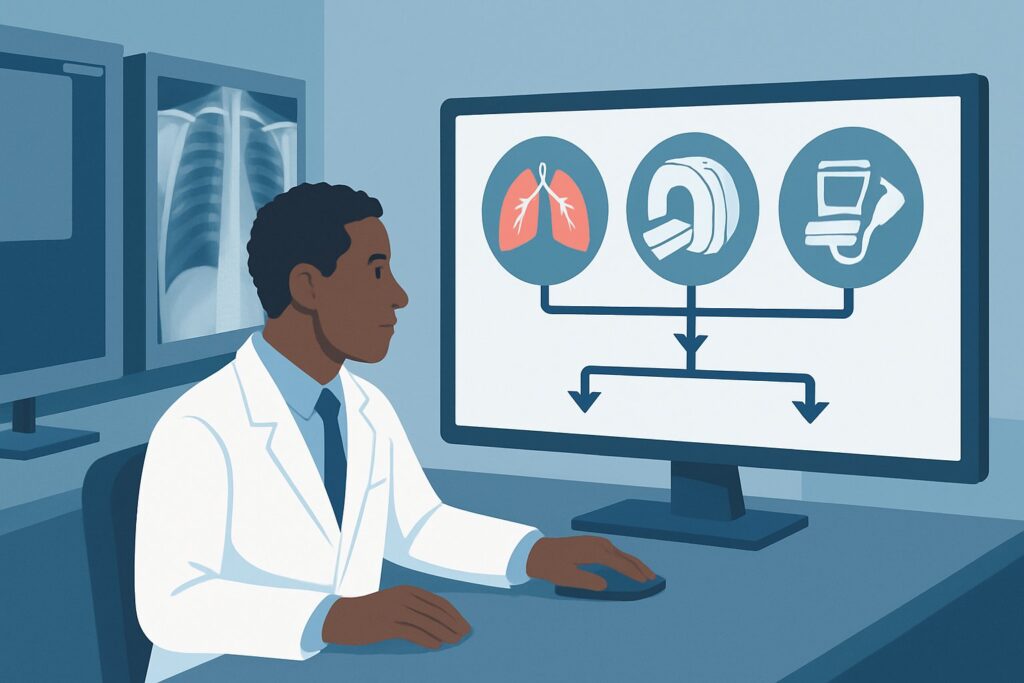
It’s 4 PM on a Tuesday, and you’re reviewing the results of a low-dose screening chest CT for a 68-year-old with a 40-pack-year smoking history. The report confirms a new, solid 2.2 cm nodule adjacent to the right hilum. The finding is concerning, but the location makes the next step critical. Do you watch it, attempt a needle biopsy through the chest wall, or refer for a bronchoscopic approach? This is a common and high-stakes decision point where choosing the right diagnostic procedure is key to timely diagnosis and management. This article details the American College of Radiology (ACR) Appropriateness Criteria workflow for this exact scenario: an adult smoker with a new, 1 to 3 cm solid pulmonary nodule near the hilum. For this presentation, the ACR designates Endobronchial ultrasound and biopsy as Usually Appropriate.
Who Fits This Clinical Scenario?
This guidance applies to a specific patient profile: an adult with a history of smoking who has a newly identified solitary solid pulmonary nodule measuring between 1 and 3 cm in diameter. The critical feature of this scenario is the nodule’s location—it is central, near the hilum, where the main bronchi, arteries, and veins enter the lung. This proximity to major airways and vessels is the primary determinant for the recommended diagnostic approach.
This workflow is distinct from other common pulmonary nodule presentations. This guidance does not apply if:
- The nodule is peripheral: A nodule located in the outer third of the lung parenchyma presents a different set of risks and benefits for biopsy. A serially enlarging peripheral nodule in a high-risk patient may be better suited for a different approach.
- The nodule is stable: If a solid nodule of this size has been documented as unchanged for two or more years, it is considered benign, and invasive procedures are generally not warranted.
- The nodule is ground-glass or subsolid: These nodules have a different differential diagnosis and natural history, often requiring a distinct management algorithm focused on serial imaging or specialized resectional techniques.
Correctly identifying that your patient fits this specific central, solid, new-nodule profile is the first step in applying the appropriate evidence-based pathway.
What Diagnoses Are You Working Up in This Scenario?
In an adult with a significant smoking history and a new, solid central pulmonary nodule, the primary concern is malignancy. The differential diagnosis is weighted heavily toward conditions that require definitive tissue diagnosis for treatment planning.
Primary Lung Carcinoma: This is the most significant and probable diagnosis to exclude. Non-small cell lung cancer (NSCLC), including adenocarcinoma and squamous cell carcinoma, is a leading consideration. Small cell lung cancer (SCLC) also frequently presents as a central mass and is strongly associated with smoking. The nodule’s central location and solid nature in a high-risk patient make malignancy the top differential until proven otherwise.
Carcinoid Tumor: Though less common than primary lung carcinomas, typical and atypical carcinoid tumors often arise in the central airways. They are neuroendocrine tumors that can present as well-defined hilar or perihilar nodules. While often slow-growing, they require tissue diagnosis for classification and management.
Metastatic Disease: While a solitary pulmonary nodule can be the first sign of metastatic disease from an unknown primary, it’s a less common presentation than a new primary lung cancer in a smoker. However, if the patient has a history of a non-pulmonary malignancy (e.g., renal cell carcinoma, colon cancer, melanoma), metastasis becomes a much higher consideration.
Infectious or Inflammatory Granuloma: Benign causes, such as a granuloma from a prior fungal infection (e.g., histoplasmosis) or tuberculosis, can present as a solid nodule. However, in the context of a new nodule in a high-risk smoker, these are diagnoses of exclusion that can only be confirmed after malignancy has been reasonably ruled out, typically with a biopsy.
Why Is Endobronchial Ultrasound and Biopsy the Recommended Study?
The ACR rates Endobronchial ultrasound and biopsy (EBUS) as Usually Appropriate for this scenario because it directly addresses the primary clinical question—is this malignant?—while minimizing risk given the nodule’s challenging location.
EBUS involves passing a flexible bronchoscope with an ultrasound probe at its tip into the airways. This allows for real-time visualization of the nodule and adjacent mediastinal and hilar lymph nodes through the airway wall. A fine needle can then be passed through the bronchoscope under ultrasound guidance to obtain tissue samples. This approach is highly effective for central lesions, offering high diagnostic yield for both the primary nodule and for simultaneous lymph node staging, which is critical for cancer treatment planning.
Alternative procedures are rated lower for specific reasons in this context:
- Percutaneous lung biopsy is rated May be appropriate. While effective for peripheral nodules, a percutaneous (transthoracic) needle approach to a central lesion is technically challenging and carries a higher risk of complications. Navigating past major blood vessels like the pulmonary artery and aorta, as well as the heart and main bronchi, increases the danger of hemorrhage and pneumothorax.
- Follow-up imaging only is also rated May be appropriate. This strategy of “watchful waiting” might be considered for a patient who is a very poor candidate for any procedure or who refuses invasive testing. However, for most patients, delaying diagnosis of a potential malignancy in a high-risk individual is not the preferred course, as it can allow for disease progression and loss of curative treatment options.
Procedures like surgical management or stereotactic radiotherapy are rated Usually not appropriate as a next step because they are treatments, not diagnostic tools. A tissue diagnosis is required before committing a patient to the risks of surgery or radiation.
What’s Next After Endobronchial Ultrasound and Biopsy? Downstream Workflow
The results of the EBUS-guided biopsy will direct the subsequent management pathway. The goal of the procedure is to obtain a definitive diagnosis that allows the clinical team to move forward with confidence.
- If the biopsy is positive for malignancy: This result triggers a full staging workup and multidisciplinary consultation. The patient will typically undergo a PET/CT scan to assess for metastatic disease and a brain MRI, particularly if small cell lung cancer is diagnosed. The case should then be presented at a multidisciplinary tumor board including thoracic surgery, medical oncology, and radiation oncology to determine the optimal treatment plan based on the cancer type, stage, and the patient’s overall health.
- If the biopsy is negative for malignancy (e.g., shows inflammation or normal lung tissue): This is a critical juncture. A negative result from a central nodule in a high-risk patient must be interpreted with caution. If the clinical suspicion for cancer remains high, the result may be considered a false negative or non-diagnostic. The next step often involves discussion with the multidisciplinary team. Options may include proceeding to a percutaneous biopsy (if technically feasible and the risk is acceptable), surgical biopsy (e.g., mediastinoscopy or video-assisted thoracoscopic surgery), or short-interval follow-up CT imaging to assess for growth.
- If the biopsy yields a specific benign diagnosis: If the pathology confidently identifies a benign entity, such as a granuloma, further invasive workup is typically unnecessary. Management may involve a follow-up CT scan in 6-12 months to ensure stability, after which no further imaging is needed.
Pitfalls to Avoid (and When to Get Help)
Navigating the workup of a central pulmonary nodule requires careful consideration to avoid common errors. First, do not assume a non-diagnostic or negative EBUS biopsy definitively rules out cancer in a high-risk patient; the pre-test probability of malignancy remains high. Second, avoid the pitfall of ordering a PET/CT before obtaining a tissue diagnosis; while useful for staging, many inflammatory conditions can be PET-avid, leading to false positives. Finally, be cautious about attributing a central lesion to a vascular cause without definitive imaging; ensure the finding is a true nodule and not an aneurysm or other vascular anomaly. If the EBUS is non-diagnostic and clinical suspicion is high, escalate the case to a multidisciplinary tumor board for consensus on the next best step, which may include surgical intervention.
Related ACR Topics and Tools
The workup of pulmonary nodules is complex, with guidance varying based on nodule size, characteristics, and patient risk factors. For a comprehensive overview of all clinical variants, it is helpful to consult the parent topic guide and utilize available decision-support resources.
- For breadth across all scenarios in Radiologic Management of Pulmonary Nodules and Masses, see our parent guide: Radiologic Management of Pulmonary Nodules and Masses: ACR Appropriateness Decoded.
- To explore other clinical scenarios and their corresponding ACR ratings, use the ACR Appropriateness Criteria Lookup.
- For details on imaging techniques and protocols for various studies, refer to the Imaging Protocol Library.
- To discuss cumulative radiation exposure with patients who have undergone multiple CT scans, the Radiation Dose Calculator can be a useful tool.
Frequently Asked Questions
Why not order a PET/CT scan first to see if the nodule is ‘hot’?
While PET/CT is essential for staging a diagnosed cancer, it is not the recommended first diagnostic step for the nodule itself. Many benign inflammatory or infectious conditions, such as granulomas, can also be metabolically active and appear ‘hot’ on a PET scan, leading to a false-positive result. A tissue biopsy provides a definitive pathologic diagnosis, which is required before initiating treatment. The ACR guidelines prioritize obtaining a tissue diagnosis first.
What technically defines a nodule as ‘hilar’ or ‘central’?
Generally, a central or hilar nodule is located in the inner one-third to one-half of the lung, in close proximity to the main bronchi, pulmonary arteries, and veins. These are lesions that are often directly accessible or adjacent to the airways reachable by a bronchoscope, making them ideal targets for an endobronchial approach like EBUS.
Is EBUS a safe procedure for a patient with severe COPD?
EBUS is considered a minimally invasive procedure and is generally well-tolerated, even in patients with underlying lung disease like Chronic Obstructive Pulmonary Disease (COPD). It avoids the risk of pneumothorax associated with percutaneous biopsies. However, any procedure involving sedation and airway instrumentation carries some risk. The decision should be made after a careful risk/benefit assessment by the interventional pulmonologist or thoracic surgeon, often including pre-procedure pulmonary function tests.
What if the nodule is slightly larger than 3 cm?
A solid lesion larger than 3 cm is technically classified as a ‘mass’ rather than a ‘nodule.’ While the principles of this guidance still largely apply (i.e., a central mass in a smoker requires a tissue diagnosis), the pre-test probability of malignancy is even higher. EBUS remains an excellent first-line diagnostic tool for central masses. The urgency for diagnosis is heightened, and the management pathway proceeds similarly.
If the EBUS biopsy comes back as non-diagnostic, what is the most common next step?
A non-diagnostic EBUS result in a high-risk patient requires further action, as the possibility of malignancy remains high. The case should be discussed at a multidisciplinary tumor board. The next step depends on the nodule’s exact location and patient fitness. Options include repeating the EBUS, attempting a percutaneous (transthoracic) needle biopsy if the anatomy is favorable, or proceeding to a surgical biopsy, such as via video-assisted thoracoscopic surgery (VATS) or mediastinoscopy, to obtain a definitive diagnosis.
Reviewed by Pouyan Golshani, MD, Interventional Radiologist — May 30, 2026