Why Are Radiographs the First Step for Clinically Suspected Osteonecrosis?
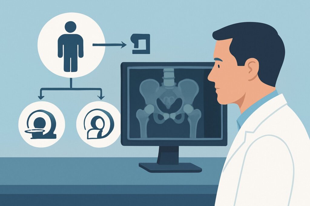
A 45-year-old man with a history of long-term corticosteroid use for lupus presents with six weeks of progressive, deep, throbbing left hip pain. The pain is worse with weight-bearing and is not improving with rest or NSAIDs. There is no history of trauma. You suspect osteonecrosis (ON) of the femoral head, but the differential is broad. Your immediate question is what imaging to order first to confirm or exclude this diagnosis without unnecessary tests or radiation. This article provides a step-by-step clinical workflow for this exact scenario, based on the American College of Radiology (ACR) Appropriateness Criteria, which rate an initial `Radiography area of interest` as Usually appropriate.
Who Fits This Clinical Scenario?
This guidance applies to patients presenting with the initial clinical suspicion of osteonecrosis, also known as avascular necrosis (AVN), prior to any imaging being performed for this specific complaint. The typical patient has new-onset, non-traumatic, focal joint pain, most commonly in the hip, but also possible in the knee, shoulder, or ankle. A key component is the presence of one or more significant risk factors, such as:
- Systemic corticosteroid use (the most common non-traumatic cause)
- Excessive alcohol consumption
- Sickle cell disease or other hemoglobinopathies
- Systemic lupus erythematosus (SLE)
- History of organ transplantation
- Gaucher disease
- Caisson disease (decompression sickness)
This workflow is specifically for the initial diagnostic step. It does not apply to patients who have already undergone imaging for this problem. For instance, if a patient with these symptoms has already had normal radiographs, they fit a different clinical scenario requiring a different imaging approach. Similarly, this guidance is not for patients with known osteonecrosis who need follow-up or pre-surgical planning, as those represent distinct clinical questions with their own recommended imaging pathways.
What Diagnoses Are You Working Up in This Scenario?
When you order the initial radiograph for suspected osteonecrosis, you are evaluating a differential diagnosis that extends beyond just ON. The goal of the first imaging study is to identify classic signs of ON or point toward an alternative diagnosis.
Osteonecrosis (Avascular Necrosis): This is the primary concern. The pathology involves the death of bone tissue due to a loss of blood supply. Early in the disease, radiographs may be normal, but as it progresses, findings like sclerosis, subchondral lucency (the crescent sign), and eventually femoral head flattening and collapse can become apparent. Identifying these signs confirms the diagnosis.
Subchondral Insufficiency Fracture: This can present with acute pain similar to ON, particularly in older patients with osteoporosis or in younger, active individuals. Radiographs may show a subchondral fracture line or sclerosis, which can sometimes mimic the findings of osteonecrosis, although the clinical context often helps differentiate them.
Degenerative Osteoarthritis: Especially in the hip or knee, osteoarthritis is a common cause of joint pain. Radiographs are excellent for identifying characteristic findings like joint space narrowing, osteophyte formation, and subchondral sclerosis, which are distinct from the typical early signs of ON.
Transient Osteoporosis of the Hip (TOH): A less common, self-limiting condition that causes bone marrow edema and significant pain. Radiographs in TOH are often normal initially but may later show diffuse osteopenia around the joint without the focal collapse seen in ON. It is a diagnosis of exclusion, often confirmed on MRI.
Why Is Radiography the Recommended Initial Study for This Presentation?
For the initial evaluation of suspected osteonecrosis, the ACR designates `Radiography area of interest` as Usually appropriate. This recommendation is based on a combination of diagnostic utility, accessibility, and cost-effectiveness. Radiographs serve as a crucial first-line screening tool.
The primary strength of radiography is its ability to either establish the diagnosis or suggest an alternative. If characteristic findings of moderate to advanced ON are present—such as the pathognomonic “crescent sign” (a subchondral lucency), sclerosis, or articular surface collapse—the diagnosis is confirmed, and the subsequent workup can proceed directly to staging and treatment planning. Furthermore, radiographs are highly effective at identifying other common causes of joint pain, like advanced osteoarthritis or obvious fractures, which may resolve the clinical question immediately.
Conversely, other powerful imaging modalities are rated lower for this specific initial step:
- MRI area of interest without IV contrast is rated Usually not appropriate for the initial workup. While MRI is the most sensitive and specific test for detecting early-stage ON (before radiographic changes appear), ordering it as the first test is not the most efficient pathway. It is more resource-intensive and is best reserved for cases where radiographs are negative or equivocal but clinical suspicion remains high.
- Bone scan area of interest is also rated Usually not appropriate. This nuclear medicine study has a high radiation dose (☢☢☢ 1-10 mSv) and suffers from low specificity. It can show increased radiotracer uptake in the affected area, but this finding is non-specific and can be seen in fractures, infection, arthritis, and ON, failing to provide a definitive diagnosis.
Radiography provides a low-radiation (RRL=Varies), low-cost, and readily available method to triage the patient. It effectively sorts patients into three bins: those with clear ON, those with a clear alternative diagnosis, and those with normal or equivocal findings who require further investigation.
What’s Next After Radiography? Downstream Workflow
The results of the initial radiograph directly guide your next steps. The clinical pathway diverges significantly depending on the findings, highlighting the radiograph’s role as a critical decision-making node.
If the radiograph is positive for osteonecrosis: When findings like a crescent sign, femoral head flattening, or significant sclerosis are identified, the diagnosis is made. The next step is typically to stage the disease and assess the extent of involvement. This often involves ordering an MRI to evaluate the size of the necrotic lesion, check for bilaterality (as ON is frequently bilateral, especially in the hips), and determine if the articular cartilage is intact. An orthopedic surgery consultation is warranted for management, which may range from observation to core decompression or joint arthroplasty depending on the stage.
If the radiograph is negative: A normal radiograph does not exclude early-stage osteonecrosis. If your clinical suspicion remains high based on the patient’s symptoms and risk factors, you should proceed to the next logical step in the diagnostic algorithm. This moves the patient into a different ACR scenario: “Clinically suspected osteonecrosis. Normal radiographs…” For that scenario, the recommended study is an MRI without contrast, which is highly sensitive for detecting the bone marrow edema and other early changes of ON not visible on a plain film.
If the radiograph is indeterminate: Sometimes, findings are equivocal, such as subtle sclerosis or questionable subchondral changes. In these cases, MRI is the definitive problem-solving tool. It can differentiate early ON from other conditions like a subchondral insufficiency fracture or transient osteoporosis, providing a clear diagnosis to guide further management.
Pitfalls to Avoid (and When to Get Help)
In the initial workup of suspected osteonecrosis, several common pitfalls can delay diagnosis or lead to inefficient testing. First, avoid stopping the workup after a negative radiograph in a high-risk patient with persistent, classic symptoms. Radiographs are insensitive in early disease (Ficat-Arlet stage 0 and I). Second, when imaging the hip, always consider ordering bilateral views, as osteonecrosis is bilateral in a high percentage of non-traumatic cases; identifying asymptomatic contralateral disease is clinically important. Third, do not order a CT scan or bone scan as the next step after a negative radiograph; MRI is the superior modality for assessing early-stage ON. If you are uncertain about interpreting subtle radiographic findings or if the clinical picture is complex, a consultation with a musculoskeletal radiologist or an orthopedic surgeon can provide crucial guidance.
Related ACR Topics and Tools
For a comprehensive overview of imaging recommendations across all clinical presentations of osteonecrosis, from initial suspicion to pre-operative planning, please refer to our parent guide. For tools to help you navigate other clinical scenarios or understand imaging protocols and radiation safety, the following resources are available.
- For breadth across all scenarios in Osteonecrosis, see our parent guide: Osteonecrosis: ACR Appropriateness Decoded.
- ACR Appropriateness Criteria Lookup — for adjacent scenarios
- Imaging Protocol Library — for technique on the recommended study
- Radiation Dose Calculator — for cumulative dose conversations
Frequently Asked Questions
Why not just order an MRI first, since it’s more sensitive for early osteonecrosis?
While MRI is the most sensitive test, the American College of Radiology recommends a stepwise approach for efficiency and resource stewardship. Radiographs are inexpensive, widely available, and can often confirm the diagnosis or reveal an alternative cause of pain, like osteoarthritis. MRI is best reserved as the next step for patients with negative or indeterminate radiographs but high clinical suspicion, avoiding unnecessary advanced imaging for a significant portion of patients.
Should I order bilateral hip radiographs if the pain is only on one side?
Yes, it is generally recommended to obtain bilateral views, typically an AP pelvis and frog-leg lateral views of both hips. Non-traumatic osteonecrosis is bilateral in a high percentage of cases (up to 80% in some series), and identifying asymptomatic disease in the contralateral hip is critical for comprehensive management and patient counseling.
What specific radiographic views are most helpful for suspected hip osteonecrosis?
The standard views are an anteroposterior (AP) view of the pelvis and a frog-leg lateral view of the affected hip. The AP pelvis allows for comparison with the asymptomatic side, while the frog-leg lateral view can better profile the anterosuperior aspect of the femoral head, where osteonecrosis and the associated subchondral crescent sign often first appear.
What if the patient has a contraindication to MRI, like an incompatible pacemaker?
If a patient has a strong contraindication to MRI and radiographs are negative, a CT scan or a bone scan may be considered as alternative, second-line options. While CT is less sensitive than MRI for early disease, it can detect sclerosis and subchondral fractures. A bone scan can show increased uptake but is non-specific. In such complex cases, consultation with a radiologist is highly recommended to determine the best alternative imaging strategy.
How does this initial workup change if the patient is a child with suspected Legg-Calvé-Perthes disease?
Legg-Calvé-Perthes disease is an idiopathic osteonecrosis of the femoral head in children. The initial imaging approach is the same: start with AP and frog-leg lateral radiographs of the hips. Radiographic findings guide the diagnosis and staging. MRI is used more frequently in children for early detection and to assess the extent of femoral head involvement and perfusion, which has prognostic value.
Reviewed by Pouyan Golshani, MD, Interventional Radiologist — May 30, 2026