Why Does the ACR Advise Against Imaging for High-Risk Colorectal Cancer Screening?
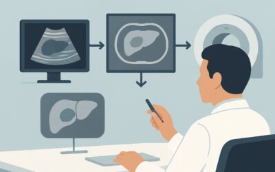
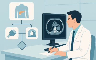
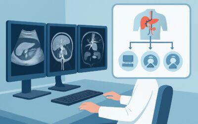
A 48-year-old man with a strong family history of colorectal cancer (CRC)—his father was diagnosed at 52 and a paternal aunt at 55—is in your office to establish a screening plan. He is asymptomatic but anxious about the invasive nature of a colonoscopy and asks if a “virtual colonoscopy” (CT colonography) is a reasonable alternative. You need to decide on the appropriate screening modality, considering his high-risk status and the specific recommendations for this patient population. This article explains the clinical workflow and imaging rationale for colorectal cancer screening in high-risk individuals. Based on the American College of Radiology (ACR) Appropriateness Criteria, imaging studies like CT colonography and Fluoroscopy barium enema double-contrast are designated as Usually not appropriate for this specific clinical scenario.
Who Qualifies as a High-Risk Individual for Colorectal Cancer Screening?
This guidance applies to asymptomatic adults who are considered high-risk for developing colorectal cancer. Defining this category is crucial, as the screening recommendations differ significantly from those for average-risk or elevated-risk individuals. A patient is generally considered high-risk if they have one or more of the following factors:
- A personal history of colorectal cancer or adenomatous polyps.
- A known or suspected hereditary colorectal cancer syndrome, such as Lynch syndrome (Hereditary Non-Polyposis Colorectal Cancer or HNPCC) or Familial Adenomatous Polyposis (FAP).
- A strong family history, which typically means a first-degree relative (parent, sibling, or child) diagnosed with CRC or an advanced adenoma before age 60, or two first-degree relatives diagnosed at any age.
- A personal history of inflammatory bowel disease (IBD), such as ulcerative colitis or Crohn’s disease, particularly with pancolitis of long duration (greater than 8-10 years).
This scenario is distinct from others. It does not apply to average-risk individuals, who have no known risk factors other than age (45 or older). It also differs from elevated-risk individuals, who may have a less significant family history (e.g., a first-degree relative diagnosed at age 60 or older). Finally, this guidance is for initial or surveillance screening, not for patients who have already undergone an incomplete optical colonoscopy, which is a separate clinical scenario with its own imaging recommendations.
What Are the Primary Targets of High-Risk Colorectal Cancer Screening?
In any screening program, the goal is to detect and address disease at its earliest, most treatable stage. For high-risk colorectal cancer screening, the primary targets are twofold: early-stage, asymptomatic colorectal adenocarcinoma and its precursor lesions, adenomatous polyps.
Adenomatous Polyps: These are the most common precursor lesions for colorectal cancer. The progression from a small adenoma to an invasive carcinoma (the adenoma-carcinoma sequence) can take several years. For high-risk individuals, particularly those with genetic syndromes, this progression may be accelerated, and the number of polyps can be far greater. The fundamental goal of screening in this population is not just to find these polyps but to remove them (polypectomy) before they can undergo malignant transformation. This “see and treat” capability is a central concept in CRC prevention.
Early-Stage Colorectal Cancer: If a cancer has already developed, detecting it when it is small, localized (Stage I or II), and has not spread to lymph nodes or distant organs dramatically improves prognosis. High-risk individuals are, by definition, more likely to develop cancer and to do so at a younger age. An effective screening strategy must have high sensitivity for detecting these early cancers, which may be subtle, flat, or located in difficult-to-visualize areas of the colon.
The clinical workup is therefore not just diagnostic but inherently therapeutic. The choice of screening modality must account for the need to both identify and remove precursor lesions in a single session if possible.
Why Is Imaging ‘Usually Not Appropriate’ for Screening High-Risk Individuals?
While imaging modalities like CT colonography are valuable in other contexts, the ACR rates them as “Usually not appropriate” for primary screening in high-risk individuals. The rationale is grounded in the unique requirements of this patient population, where the ability to perform simultaneous diagnosis and therapy is paramount.
The primary recommended screening test for high-risk individuals is optical colonoscopy. Unlike imaging, colonoscopy allows for direct visualization of the colonic mucosa, biopsy of suspicious lesions, and removal of polyps during the same procedure. This single-step approach is crucial for prevention and is the established standard of care.
Let’s examine why specific imaging studies are not recommended:
- CT Colonography (CTC): This study, also known as virtual colonoscopy, is rated Usually not appropriate. While CTC has good sensitivity for polyps larger than 10 mm, its performance for smaller or flat lesions—which can be particularly significant in patients with IBD or Lynch syndrome—is lower than that of colonoscopy. Most importantly, if CTC identifies a significant polyp, the patient must still undergo a subsequent conventional colonoscopy for removal. This creates a two-step process that increases cost, patient burden, and potential delays in treatment. The radiation dose (☢☢☢☢ 10-30 mSv) is also a consideration, especially for patients who will require frequent surveillance over many years.
- Fluoroscopy Barium Enema (Double-Contrast): This study is also rated Usually not appropriate. Its sensitivity for detecting polyps and early cancers is significantly lower than both colonoscopy and CT colonography. It is particularly poor at identifying smaller polyps and flat lesions. Given the availability of superior alternatives and a notable radiation dose (☢☢☢ 1-10 mSv), its role in modern CRC screening for any risk group, especially high-risk, is virtually nonexistent.
In summary, for high-risk patients, the clinical need extends beyond simple detection. The ability to intervene by removing precursor lesions makes optical colonoscopy the only appropriate primary screening tool. Imaging studies are relegated to a problem-solving role, such as after an incomplete colonoscopy, but not for initial screening.
What Is the Correct Downstream Workflow for a High-Risk Patient?
Once a patient is correctly identified as high-risk, the workflow bypasses screening imaging and proceeds directly to endoscopic evaluation. The key decision points revolve around the timing and frequency of this surveillance.
Initial Step: Referral to Gastroenterology. The first and most critical step is to refer the patient to a gastroenterologist for consultation and to schedule a high-quality optical colonoscopy. Genetic counseling may also be appropriate if a hereditary syndrome is suspected based on personal or family history.
If Colonoscopy is Normal: A “normal” result is reassuring, but it does not change the patient’s high-risk status. Surveillance intervals are much shorter than for the general population. For example:
- Individuals with Lynch syndrome may require colonoscopy every 1-2 years, starting in their 20s.
- Those with a first-degree relative diagnosed with CRC before age 60 are typically advised to begin screening 10 years prior to the age of the relative’s diagnosis, or at age 40, whichever is earlier, with repeat colonoscopy every 5 years.
- Patients with long-standing IBD require surveillance colonoscopy with multiple biopsies every 1-3 years.
These intervals are guided by specific societal guidelines (e.g., from the American Gastroenterological Association or the U.S. Multi-Society Task Force on Colorectal Cancer).
If Polyps are Found: The downstream workflow depends on the number, size, and pathology of the polyps removed. The presence of high-risk adenomas (e.g., large size, villous features, high-grade dysplasia) will necessitate a shorter follow-up interval, often in 1 to 3 years, to ensure complete clearance and monitor for new lesions.
Pitfalls to Avoid (and When to Get Help)
Managing high-risk individuals for colorectal cancer screening requires careful risk stratification and adherence to evidence-based pathways. Several common pitfalls can compromise patient care.
A primary pitfall is ordering a non-invasive imaging test like CT colonography as a “compromise” for a patient hesitant to undergo colonoscopy. This delays definitive management, as a positive finding will require a colonoscopy anyway. Another error is miscalculating the screening start date or surveillance interval by not fully appreciating the significance of the family history (e.g., the age of a relative’s diagnosis). Finally, failing to refer for genetic counseling when a hereditary syndrome is suspected can miss an opportunity to identify at-risk family members. If you are uncertain about a patient’s risk category or the appropriate surveillance schedule, a consultation with a gastroenterologist is the most appropriate next step.
Related ACR Topics and Tools
For a comprehensive overview of imaging across all risk profiles and clinical presentations, please see our parent guide. Additional GigHz tools can help you navigate adjacent scenarios, understand imaging techniques, and discuss radiation safety with your patients.
- For breadth across all scenarios in Colorectal Cancer Screening, see our parent guide: Colorectal Cancer Screening: ACR Appropriateness Decoded.
- To look up appropriateness criteria for other clinical scenarios: ACR Appropriateness Criteria Lookup.
- To review technical details for various imaging studies: Imaging Protocol Library.
- To help frame conversations about medical radiation with patients: Radiation Dose Calculator.
Frequently Asked Questions
What if my high-risk patient absolutely refuses a colonoscopy?
This is a challenging clinical situation that requires shared decision-making. While imaging is rated ‘Usually not appropriate’ for screening, if a patient refuses the standard of care, a test like CT colonography may be considered better than no screening at all. However, it’s critical to counsel the patient that any significant finding on the CT will still require a follow-up colonoscopy for confirmation and removal.
What defines a ‘strong family history’ that places someone in the high-risk category?
Generally, a high-risk family history includes having a first-degree relative (parent, sibling, child) diagnosed with colorectal cancer or an advanced adenoma before age 60, or having two or more first-degree relatives diagnosed at any age. This is distinct from an elevated-risk history, such as having one first-degree relative diagnosed at age 60 or older.
Is there any role for imaging in a high-risk patient with inflammatory bowel disease (IBD)?
For screening and surveillance for dysplasia in IBD, optical colonoscopy with biopsies is the standard. However, imaging such as CT enterography or MR enterography plays a crucial role in assessing disease activity, extent, and complications of IBD (like strictures or fistulas), but it is not used for routine cancer screening in this population.
Why is the radiation from CT colonography a particular concern for high-risk patients?
High-risk patients often begin screening at a younger age and require surveillance much more frequently than the general population (e.g., every 1-5 years). The cumulative radiation exposure from repeated CT scans over a lifetime could become significant, which is another reason why a non-radiation-based modality like colonoscopy is strongly preferred.
If a patient has a known genetic syndrome like FAP, is imaging ever used?
For patients with Familial Adenomatous Polyposis (FAP), the standard is frequent endoscopic surveillance of the colon and rectum (and upper GI tract). Imaging is not used for screening but may be used to evaluate for extra-colonic manifestations, such as desmoid tumors, which can be assessed with CT or MRI of the abdomen and pelvis.
Reviewed by Pouyan Golshani, MD, Interventional Radiologist — May 29, 2026