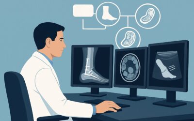
Why Is Imaging Not Recommended for Ankle Trauma with Negative Ottawa Rules?
A 28-year-old patient presents to your urgent care clinic after twisting their ankle during a weekend soccer game. They are ambulatory, though with a limp, and your physical examination is reassuring. There is no bony tenderness over the posterior edge or tip of either malleolus, nor is there tenderness over the navicular or the base of the fifth metatarsal. The patient can bear weight for four steps in the clinic. According to the Ottawa Ankle Rules, the pre-test probability for a clinically significant fracture is exceedingly low. This article addresses the specific American College of Radiology (ACR) guidance for this common scenario: an adult or child over 5 with acute ankle trauma who is negative by the Ottawa Ankle Rules. For this presentation, initial imaging studies, including radiography and ultrasound, are rated as Usually Not Appropriate.
Who Fits This Clinical Scenario?
This guidance applies to a well-defined patient population to maximize safety and avoid unnecessary testing. The key inclusion criteria are:
- An adult or child aged 5 years or older.
- History of acute ankle trauma (typically within 48 hours).
- The patient is neurologically intact, with no peripheral neuropathy or other conditions that would confound a physical examination.
- The patient meets the criteria for evaluation by the Ottawa Ankle Rules (OAR), and the results are negative.
A negative OAR exam means the patient has no bony point tenderness in the malleolar zones or midfoot zones and is able to bear weight immediately after the injury and for four steps at the time of evaluation. This clinical decision rule has a very high sensitivity for excluding clinically significant ankle and midfoot fractures.
This workflow is distinct from other clinical situations. If a patient has persistent pain for more than a week despite conservative management, or if they have positive findings on their OAR exam (e.g., point tenderness over the lateral malleolus), they fall into a different category where imaging is often warranted. Similarly, patients with overlying skin breakdown, suspected open fracture, or neurologic compromise are excluded from this specific low-risk pathway.
What Diagnoses Are You Working Up in This Scenario?
When the Ottawa Ankle Rules are negative, a clinically significant fracture is effectively ruled out, and the diagnostic focus shifts to soft tissue injuries. The goal of withholding imaging is not to ignore the patient’s pain but to recognize that the likely diagnoses are self-limiting conditions that are managed clinically, without the need for radiographic confirmation.
Ankle Sprain (Grade I or II): This is the most common diagnosis. It involves stretching or partial tearing of the ankle ligaments, most frequently the anterior talofibular ligament (ATFL). The patient will have localized swelling and pain, but the absence of specific bony tenderness and the ability to bear weight point away from a severe, unstable injury or associated fracture that would alter initial management.
Soft Tissue Contusion: A direct blow or forceful twisting can cause a deep bruise to the muscles, fat, and skin around the ankle without significant ligamentous injury. This can be quite painful and cause swelling but resolves with conservative care. Imaging would show non-specific soft tissue swelling and edema but would not change the treatment plan.
Mild Peroneal Tendinopathy or Tenosynovitis: The injury mechanism can sometimes strain the peroneal tendons running along the outside of the ankle. While more significant tendon tears or dislocations can occur, in a low-risk OAR-negative patient, mild inflammation or strain is more likely. This is initially managed identically to an ankle sprain.
Why Is Imaging Usually Not Appropriate for This Presentation?
The ACR Appropriateness Criteria panel designates all initial imaging modalities as “Usually Not Appropriate” for this specific clinical scenario, a recommendation rooted in the high performance of the Ottawa Ankle Rules and the principles of evidence-based medicine.
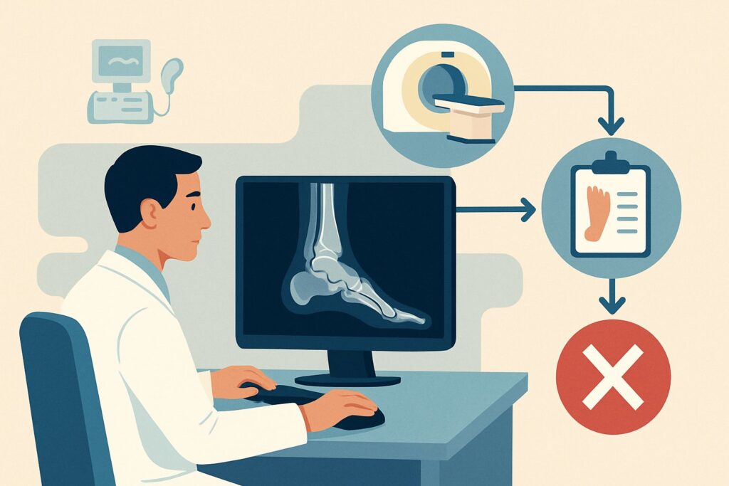
The primary rationale is the exceptionally high negative predictive value of the OAR, which approaches 100% for excluding significant malleolar, talar, or calcaneal fractures. When a validated clinical decision rule can so effectively rule out the primary diagnosis that would require a change in management (i.e., immobilization or surgical consultation for a fracture), performing imaging offers little to no clinical benefit. It exposes the patient to unnecessary costs and, in the case of radiography or CT, ionizing radiation, without altering the initial treatment plan of symptomatic care.
Let’s review the specific modalities and their ratings:
- Radiography ankle is rated Usually Not Appropriate. The yield for finding a fracture in this OAR-negative population is extremely low. While the radiation dose is small (less than 0.1 mSv for an adult), the ACR guidance supports avoiding any unnecessary radiation exposure when the clinical benefit is negligible.
- US ankle is also rated Usually Not Appropriate for initial evaluation. While ultrasound is excellent for evaluating ligaments and tendons and involves no radiation (0 mSv), these findings do not change acute management. A Grade I sprain and a Grade II sprain are both treated with rest, ice, compression, and elevation (RICE). Identifying the specific torn ligament fibers on an initial ultrasound is academic and does not improve the patient’s outcome.
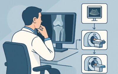
- MRI ankle without IV contrast is similarly rated Usually Not Appropriate. MRI provides exquisite detail of soft tissues but is a high-cost, resource-intensive study reserved for cases of persistent pain, instability, or suspicion of a more complex injury (like an osteochondral lesion) after a trial of conservative therapy has failed—a different clinical scenario entirely.
What’s Next? Downstream Workflow After a Negative OAR Exam
With imaging deferred, the clinical workflow focuses on patient education, symptomatic management, and establishing clear follow-up criteria. This approach empowers the patient while ensuring a safety net is in place.
If the exam is consistent with a low-grade sprain or contusion (the expected finding): The appropriate next step is a course of conservative management. This typically includes the RICE protocol, activity modification, and over-the-counter analgesics like NSAIDs if not contraindicated. Provide the patient with clear “return to clinic” instructions: if they are unable to bear weight after 3-4 days, if the pain significantly worsens, or if the swelling does not begin to improve within 5-7 days, they should be re-evaluated. At that point, they may fit a different clinical scenario, such as acute trauma to the ankle with persistent pain, where imaging may become appropriate.
If the patient develops new symptoms: The onset of numbness, tingling, skin discoloration, or coldness in the foot are red flags that warrant immediate re-evaluation to assess for neurovascular compromise or compartment syndrome, though these are rare in this low-risk injury pattern.
The key is to treat the patient, not the image. For an OAR-negative ankle injury, the treatment is clinical, and the downstream workflow involves monitoring the expected clinical improvement.
Pitfalls to Avoid (and When to Get Help)
Several common pitfalls can occur in managing these seemingly straightforward injuries. First, ensure you are applying the Ottawa Ankle Rules correctly; they are only validated for non-intoxicated, neurologically intact patients who can cooperate with an exam. Second, avoid ordering imaging simply due to patient anxiety or for “reassurance.” A confident explanation of why imaging is not needed is a cornerstone of good stewardship. Third, do not neglect to provide specific, time-based follow-up instructions. Simply saying “come back if it’s not better” is too vague. If the patient’s pain is out of proportion to the physical exam findings or fails to improve as expected within a week, it is time to escalate care, which may include re-examination and proceeding with imaging.
Related ACR Topics and Tools
This article covers one specific variant within the broader topic of acute ankle trauma. For a comprehensive overview of all related scenarios, from suspected syndesmotic injury to post-operative pain, please consult our parent guide. The tools below can help you apply ACR guidance to your daily practice.
- For breadth across all scenarios in Acute Trauma to the Ankle, see our parent guide: Acute Trauma to the Ankle: ACR Appropriateness Decoded.
- To look up other clinical scenarios, use the Imaging Appropriateness Selector.
- To review imaging techniques for studies you do order, see the Imaging Protocol Library.
- To discuss radiation exposure with patients, consult the Radiation Dose Calculator.
Frequently Asked Questions
Why not get an X-ray just to be 100% sure there’s no fracture?
The Ottawa Ankle Rules were specifically designed and validated to be a highly sensitive screening tool. When the rules are negative, the probability of a clinically significant ankle or midfoot fracture is less than 1%. Ordering an X-ray in this situation provides minimal additional diagnostic information while exposing the patient to unnecessary radiation and healthcare costs. The ACR guidance of ‘Usually Not Appropriate’ reflects this high level of clinical certainty.
What if the patient is an athlete and wants to know the exact grade of their ligament sprain?
In the acute setting for an OAR-negative injury, the initial management for a Grade I (stretching) and Grade II (partial tear) ligament sprain is identical: rest, ice, compression, elevation, and gradual return to activity. While an ultrasound or MRI could differentiate these, it does not change the immediate treatment plan. Advanced imaging is typically reserved for cases of chronic instability or if the injury fails to improve after an appropriate course of conservative therapy and rehabilitation.
Does this guidance apply to children under 5 years old?
No, this specific ACR variant and the Ottawa Ankle Rules are intended for patients aged 5 and older. Younger children can have different injury patterns (e.g., physeal fractures) and may be unable to cooperate fully with the physical exam required for the OAR. Evaluating acute ankle trauma in very young children requires a different clinical approach and a lower threshold for ordering radiographs.
If I don’t order imaging, what should I document to support my decision?
Excellent documentation is key. Your note should explicitly state that the patient was evaluated using the Ottawa Ankle Rules and that the findings were negative. Document the specific negative findings: no bony tenderness at the four key locations (posterior edge/tip of medial and lateral malleoli, navicular, base of 5th metatarsal) and the patient’s ability to bear weight for at least four steps. This documentation clearly justifies the decision to defer imaging based on a validated clinical decision rule.
What if the patient has significant swelling but is still OAR-negative?
The degree of swelling does not override a negative Ottawa Ankle Rules exam. Significant soft tissue swelling can occur with ligamentous sprains and contusions in the absence of a fracture. The management remains focused on conservative care (RICE protocol). However, if the swelling is rapidly progressive or associated with tense, woody compartments and pain out of proportion, you should have a high suspicion for compartment syndrome, which is a clinical diagnosis and a surgical emergency.
Reviewed by Pouyan Golshani, MD, Interventional Radiologist — May 26, 2026