What Imaging Should You Order for Suspected Pseudogout When Radiographs Are Normal?
A 68-year-old male presents with a six-month history of intermittent, sharp pain and swelling in his right knee. The episodes are self-limiting but are becoming more frequent. You suspect a crystalline arthropathy, specifically calcium pyrophosphate dihydrate (CPPD) disease, often called pseudogout. You order initial radiographs of the knee, which return showing only mild degenerative changes, with no definitive chondrocalcinosis. The clinical picture strongly suggests CPPD, but the first-line imaging is inconclusive. What is the most appropriate next step to confirm the diagnosis and guide management? This article provides a detailed workflow for this specific clinical scenario, based on the American College of Radiology (ACR) Appropriateness Criteria, which rate a `US area of interest` as “Usually Appropriate.”
Who Fits This Clinical Scenario for Chronic Joint Pain?
This guidance is intended for a specific patient population: adults with chronic or recurrent extremity joint pain where there is a moderate to high clinical suspicion for calcium pyrophosphate dihydrate (CPPD) disease. The key feature of this scenario is that initial radiographs of the affected joint have already been performed and were either normal, non-diagnostic, or showed inconclusive findings like mild osteoarthritis without classic chondrocalcinosis.
Inclusion Criteria:
- Chronic (weeks to months) or episodic mono- or oligoarticular joint pain.
- Clinical features suggestive of CPPD, such as acute inflammatory flares mimicking gout, affecting joints like the knee, wrist, or shoulder.
- Initial radiographs are complete but do not confirm the diagnosis.
Exclusion Criteria (These patients follow a different workflow):
- High Suspicion for Gout: If the patient has a history of hyperuricemia and podagra (pain in the first metatarsophalangeal joint), the workup shifts. See the specific guidance for suspected gout with normal radiographs.
- High Suspicion for Inflammatory Arthritis: If the patient presents with symmetric polyarthritis, significant morning stiffness, and positive serologies (e.g., RF, anti-CCP), the diagnostic pathway for inflammatory arthritis should be followed.
- Definitive Radiographic Findings: If initial radiographs clearly show chondrocalcinosis in the fibrocartilage or hyaline cartilage, further imaging to confirm CPPD is often unnecessary.
What Diagnoses Are You Working Up in This Scenario?
When initial radiographs are unrevealing in a patient with suspected CPPD, advanced imaging aims to differentiate between several overlapping conditions. The goal is to visualize the specific pathologic features that plain films may miss.
Calcium Pyrophosphate Dihydrate (CPPD) Disease: This is the primary diagnosis of concern. The condition is caused by the deposition of calcium pyrophosphate crystals in and around joints. Imaging is tasked with directly visualizing these crystal deposits, which manifest as chondrocalcinosis (calcification of cartilage). Advanced imaging can detect these deposits with higher sensitivity than radiography, especially when the burden is low or in locations obscured on plain film.
Gout (Monosodium Urate Crystal Deposition Disease): Gout is the most common crystalline arthropathy and a key mimicker of CPPD. Clinically, the acute flares can be indistinguishable. While radiographs are often normal early on, advanced imaging can detect the characteristic findings of gout, such as tophi (collections of urate crystals) and the “double contour sign” on ultrasound, which are distinct from the findings of CPPD.
Osteoarthritis (OA) with Atypical Features: CPPD can cause a secondary, destructive form of osteoarthritis. In this scenario, the goal is to determine if an underlying crystalline process is accelerating the degenerative changes seen on inconclusive radiographs. Identifying crystal deposition can change the management approach from standard OA treatment.
Inflammatory Synovitis: Less commonly, a persistent monoarthritis could represent an atypical presentation of a seronegative inflammatory arthritis. Advanced imaging can assess for synovitis, tenosynovitis, and erosions, which, while not specific, can help steer the workup toward or away from a primary inflammatory condition if no crystal deposits are found.
Why Is Ultrasound of the Area of Interest the Recommended Study?
When radiographs are inconclusive for suspected CPPD, the ACR designates `US area of interest` as a “Usually Appropriate” next step. This recommendation is based on its high diagnostic accuracy for crystalline disease, lack of ionizing radiation, and ability to provide dynamic, real-time assessment.
Ultrasound excels at visualizing the key sonographic features of CPPD with high sensitivity. The primary finding is chondrocalcinosis, which appears as a thin, hyperechoic (bright) line parallel to the surface of the hyaline cartilage or as punctate or linear hyperechoic deposits within fibrocartilage, such as the meniscus. This is distinct from the “double contour sign” of gout, where urate crystals deposit on the cartilage surface. Ultrasound can also identify associated findings like synovial hypertrophy, joint effusions, and soft tissue calcifications that may not be apparent on radiographs.
Another key advantage is its safety profile. Ultrasound uses no ionizing radiation (0 mSv), making it ideal for repeat examinations if needed. It also avoids the need for intravenous contrast agents.
Why are other studies rated lower for this specific scenario?
- CT area of interest without IV contrast is also rated “Usually Appropriate” and is highly sensitive for detecting calcification. However, it involves ionizing radiation (dose varies) and is less effective than ultrasound for evaluating synovial inflammation and soft tissue structures. It is a strong alternative if ultrasound is unavailable or if the joint is difficult to visualize sonographically.
- MRI area of interest without IV contrast is rated “Usually Not Appropriate.” While MRI is unparalleled for evaluating soft tissues, bone marrow, and cartilage morphology, it is significantly less sensitive than ultrasound or CT for detecting the fine calcifications characteristic of CPPD. The crystal deposits are often not visible on standard MRI sequences.
- Bone scan whole body is also “Usually Not Appropriate.” It is a non-specific study that detects areas of increased bone turnover. While it may be positive in an inflamed joint, the findings are not specific for CPPD and cannot differentiate it from other causes of arthritis, making it a low-yield test in this context. It also involves significant radiation exposure (☢☢☢ 1-10 mSv).
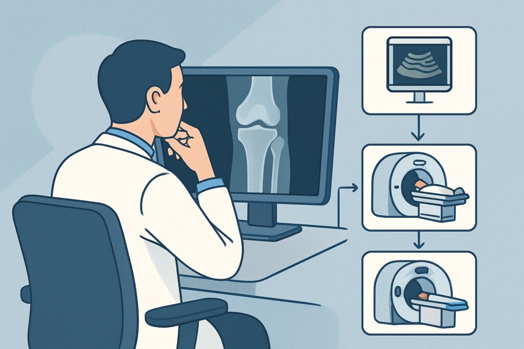
What’s Next After Ultrasound? Downstream Workflow
The results of the ultrasound will guide your subsequent clinical decisions. The workflow typically branches into three paths depending on the findings.
If the ultrasound is positive for CPPD: A definitive finding of chondrocalcinosis on ultrasound confirms the diagnosis of CPPD. The next step is clinical management, which may include joint aspiration to rule out septic arthritis during an acute flare, intra-articular corticosteroid injections, and systemic anti-inflammatory medications (e.g., NSAIDs, colchicine, or oral steroids). No further diagnostic imaging is typically required.
If the ultrasound is negative for CPPD but shows alternative findings: The sonographer may identify features suggestive of a different diagnosis. For example, the presence of a double contour sign and/or tophaceous material would strongly suggest gout, prompting a switch in management and potentially serum uric acid testing. Findings of significant synovitis and erosions without crystal deposition would raise suspicion for an inflammatory arthritis, leading to rheumatology consultation and serologic testing.
If the ultrasound is negative or non-diagnostic: If ultrasound fails to identify any specific pathology but clinical suspicion for crystalline arthropathy remains high, two main options exist. The first is to proceed with diagnostic joint aspiration for polarized light microscopy to look for crystals—the gold standard for diagnosis. The second is to consider an alternative imaging modality. In this case, a `CT area of interest without IV contrast` (also “Usually Appropriate”) could be considered, as it offers excellent sensitivity for calcification and may detect deposits missed by ultrasound. If both advanced imaging and aspiration are negative, the focus should shift to alternative diagnoses like osteoarthritis or other non-crystalline inflammatory conditions.
Pitfalls to Avoid (and When to Get Help)
Navigating the workup for suspected CPPD requires careful interpretation of both clinical and imaging findings. Here are a few common pitfalls to avoid:
- Over-relying on normal radiographs: Radiographs have low sensitivity for chondrocalcinosis. A negative X-ray does not rule out CPPD, and high clinical suspicion should prompt the next step in imaging.
- Misinterpreting ultrasound findings: The sonographic features of CPPD and gout can be subtle. Ensure the study is performed by a sonographer and interpreted by a radiologist experienced in musculoskeletal ultrasound.
- Forgetting the gold standard: While imaging is powerful, synovial fluid analysis remains the definitive test for crystalline arthropathy. If a joint effusion is present and accessible, aspiration should be strongly considered, especially to rule out co-existing infection.
- Attributing all symptoms to CPPD: Patients with CPPD often have co-existing osteoarthritis. It is important to correlate imaging findings with the patient’s specific symptoms to formulate an effective treatment plan.
If imaging is equivocal and the diagnosis remains unclear despite a thorough workup, referral to a rheumatologist is the appropriate next step for further evaluation and management.
Related ACR Topics and Tools
This article focuses on a single, specific clinical question. For a broader overview of imaging for chronic joint pain and to explore adjacent clinical scenarios, please consult our other resources. For breadth across all scenarios in Chronic Extremity Joint Pain-Suspected Inflammatory Arthritis, Crystalline Arthritis, or Erosive Osteoarthritis, see our parent guide: Chronic Extremity Joint Pain-Suspected Inflammatory Arthritis, Crystalline Arthritis, or Erosive Osteoarthritis: ACR Appropriateness Decoded.
You can also use these GigHz tools to refine your imaging orders:
- ACR Appropriateness Criteria Lookup — for adjacent scenarios
- Imaging Protocol Library — for technique on the recommended study
- Radiation Dose Calculator — for cumulative dose conversations
Frequently Asked Questions
Why is ultrasound preferred over CT when both are ‘Usually Appropriate’ for suspected CPPD?
Ultrasound is often preferred as the initial advanced imaging study because it involves no ionizing radiation, is typically more accessible and less costly, and provides superior evaluation of soft tissue inflammation (synovitis) and joint effusions. CT is an excellent alternative, particularly if ultrasound is unavailable or the anatomy is complex, but it does expose the patient to radiation.
Can I just order an MRI if I suspect joint pain and want to see everything?
While MRI provides excellent detail of soft tissues, ligaments, and bone marrow, it is rated ‘Usually Not Appropriate’ for the primary diagnosis of CPPD because it is not sensitive for detecting the calcium pyrophosphate crystal deposits. Ordering an MRI in this specific scenario is a low-yield strategy for confirming CPPD and may miss the diagnosis entirely.
What if the patient has pain in multiple joints? Should I order ultrasound of all of them?
The ACR recommends ‘US area of interest,’ meaning the study should be focused on the most symptomatic or clinically relevant joint. If CPPD is confirmed in one joint, it is considered a systemic diagnosis, and it is generally not necessary to image every painful joint unless there is a specific question, such as ruling out a septic joint in a different location.
Is joint aspiration still necessary if the ultrasound is positive for chondrocalcinosis?
For diagnostic purposes, a classic ultrasound finding of chondrocalcinosis in the right clinical context is often sufficient to confirm CPPD. However, if a patient presents with an acute, severe monoarticular flare (hot, swollen joint), joint aspiration is crucial to rule out a superimposed septic arthritis, which is a medical emergency and can coexist with a crystalline arthropathy.
Does the absence of chondrocalcinosis on ultrasound definitively rule out CPPD?
Ultrasound is highly sensitive, but not perfect. A negative ultrasound significantly lowers the probability of CPPD, but it does not completely rule it out, especially if the crystal burden is very low. If clinical suspicion remains very high despite a negative ultrasound, the next steps would be joint aspiration for microscopy (if an effusion is present) or considering a non-contrast CT.
Reviewed by Pouyan Golshani, MD, Interventional Radiologist — May 30, 2026