When to Order Imaging for Renal Transplant Dysfunction: ACR Appropriateness Decoded

It’s late in the evening, and you’re evaluating a patient with a history of renal transplant who now presents with a rising creatinine and oliguria. The differential for renal transplant dysfunction is broad, spanning from acute rejection and acute tubular necrosis (ATN) to vascular complications like stenosis or thrombosis, and urologic issues like obstruction or fluid collections. Your immediate decision is which imaging study to order first. While ultrasound is often the initial step, knowing the subsequent, more specific imaging pathways is critical for efficient and accurate diagnosis. This guide decodes the American College of Radiology (ACR) Appropriateness Criteria to help you select the right study at the right time for suspected renal transplant dysfunction.
What Does ACR Renal Transplant Dysfunction Cover?
The ACR Appropriateness Criteria for Renal Transplant Dysfunction focus specifically on the diagnostic imaging workup for an adult patient presenting with clinical or laboratory signs of a failing renal allograft. This includes scenarios where there is a new or worsening elevation in serum creatinine, a decrease in urine output, or other symptoms suggestive of graft failure. The guidelines provide a structured, evidence-based approach starting with initial imaging and branching into subsequent steps based on those initial findings.
These criteria are designed to evaluate for surgically correctable causes of graft dysfunction, such as vascular compromise (e.g., renal artery stenosis, renal vein thrombosis), urologic obstruction, or peritransplant fluid collections (e.g., hematoma, urinoma, lymphocele, abscess). The guidelines do not cover routine surveillance imaging in an asymptomatic transplant recipient or the initial postoperative assessment in an uncomplicated course. They are intended for the specific clinical problem of a newly dysfunctional allograft where imaging is needed to guide further management, which may include medical therapy, percutaneous intervention, or surgical revision.
What Imaging Should I Order for Renal Transplant Dysfunction? Recommendations by Clinical Scenario
The ACR provides a clear, stepwise approach to imaging renal transplant dysfunction, starting with a non-invasive baseline and proceeding to more advanced imaging based on the initial results. The choice of study depends heavily on the suspected etiology after the first-line test.
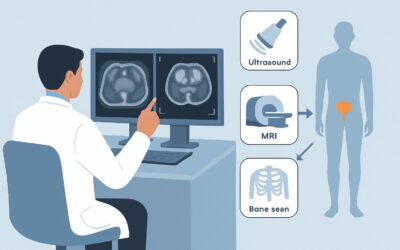
For the initial imaging of an adult with renal transplant dysfunction, the ACR panel is unequivocal. US duplex Doppler of the kidney transplant is rated as Usually appropriate. This non-ionizing, readily available modality is the cornerstone of evaluation, capable of assessing renal parenchymal echotexture, detecting hydronephrosis, identifying peritransplant fluid collections, and evaluating vascular patency and flow with Doppler. Nearly all other initial imaging modalities, including CT, MRI, and nuclear medicine scans, are rated Usually not appropriate as a first step.
If the initial ultrasound is suspicious for, but not conclusive for, an arterial etiology (such as renal artery stenosis), the next step is cross-sectional vascular imaging. Both MRA abdomen and pelvis without and with IV contrast and CTA abdomen and pelvis with IV contrast are rated Usually appropriate. The choice between them often depends on institutional preference, patient factors like renal function (for iodinated contrast) or contraindications to MRI, and the specific clinical question.
Similarly, if the ultrasound is suspicious for a venous etiology (like renal vein thrombosis), the guidelines recommend dedicated venography. MRV abdomen and pelvis without and with IV contrast and CTV abdomen and pelvis with IV contrast are both considered Usually appropriate to confirm or exclude venous pathology.
When the initial ultrasound suggests an extrinsic etiology, such as a large fluid collection causing mass effect, further characterization with cross-sectional imaging may be needed. In this scenario, multiple modalities including CT abdomen and pelvis (with or without IV contrast) and MRI abdomen and pelvis (with or without IV contrast) are rated as May be appropriate to better define the collection and its relationship to adjacent structures.
Finally, if the initial ultrasound is unremarkable or indeterminate and a parenchymal cause like acute rejection or ATN is suspected, the diagnostic pathway shifts away from non-invasive imaging. In this case, image-guided biopsy of the kidney is rated Usually appropriate. This is because conditions like rejection are primarily histologic diagnoses, and when vascular and obstructive causes have been reasonably excluded by ultrasound, direct tissue sampling is the definitive next step.
ACR Imaging Recommendations Table
| Clinical Scenario | Top Procedure | ACR Rating | Adult RRL | Pediatric RRL |
|---|---|---|---|---|
| Adult. Renal transplant dysfunction. Initial imaging. | US duplex Doppler kidney transplant | Usually appropriate | O 0 mSv | O 0 mSv [ped] |
| Adult. Renal transplant dysfunction. US suspicious for, but not conclusive for arterial etiology. Next imaging study. | MRA or CTA abdomen and pelvis with IV contrast | Usually appropriate | O 0 mSv (MRA) / ☢ ☢ ☢ ☢ 10-30 mSv (CTA) | O 0 mSv [ped] (MRA) |
| Adult. Renal transplant dysfunction. US suspicious for, but not conclusive for venous etiology. Next imaging study. | MRV or CTV abdomen and pelvis with IV contrast | Usually appropriate | O 0 mSv (MRV) / ☢ ☢ ☢ ☢ 10-30 mSv (CTV) | O 0 mSv [ped] (MRV) |
| Adult. Renal transplant dysfunction. US suspicious for, but not conclusive for extrinsic etiology. Next imaging study. | CT or MRI abdomen and pelvis | May be appropriate | ☢ ☢ ☢ 1-10 mSv (CT) / O 0 mSv (MRI) | ☢ ☢ ☢ ☢ 3-10 mSv [ped] (CT) / O 0 mSv [ped] (MRI) |
| Adult. Renal transplant dysfunction. US unremarkable or indeterminate. Next imaging study. | Image-guided biopsy kidney | Usually appropriate | Varies | Varies |
Adult vs. Pediatric Renal Transplant Dysfunction Imaging: Radiation Dose Tradeoffs
While the clinical approach to renal transplant dysfunction is similar in adults and children, imaging choices must always account for the ALARA (As Low As Reasonably Achievable) principle, especially in younger patients. Children have a longer life expectancy, and their developing tissues are more sensitive to the effects of ionizing radiation, making cumulative radiation dose a significant concern. The ACR guidelines reflect this by providing distinct pediatric relative radiation level (RRL) estimates.
For renal transplant evaluation, the first-line study, duplex Doppler ultrasound, is ideal for both populations as it involves no ionizing radiation (RRL of ‘O 0 mSv’). When cross-sectional imaging is required, the tradeoffs become more apparent. For example, a CT of the abdomen and pelvis that might impart a 1-10 mSv dose in an adult (☢ ☢ ☢) could deliver a 3-10 mSv dose in a child (☢ ☢ ☢ ☢), reflecting a higher relative level. This emphasizes the preference for non-ionizing alternatives like MRI/MRA/MRV whenever clinically feasible in pediatric patients. While CT may be faster and more accessible, the decision to use it in a child with a chronic condition requiring potential future imaging must be weighed carefully against the long-term risks of radiation exposure.
Imaging Protocol Details for Renal Transplant Dysfunction
Once you’ve decided on the right study based on the clinical scenario, ensuring it is performed correctly is the next critical step. The diagnostic yield of an imaging test is highly dependent on the right technique, contrast timing, and specific sequences or parameters used. Our protocol guides provide detailed, practical information for executing the studies recommended in these ACR criteria.
- US Carotid Doppler
- CT Abdomen/Pelvis Without Contrast (Renal Stone)
- Breast Biopsy (Ultrasound-Guided)
Tools to Help You Order the Right Study
Navigating complex clinical guidelines can be challenging, especially under time pressure. GigHz offers a suite of tools designed to support evidence-based clinical decision-making for imaging.
For scenarios beyond renal transplant dysfunction, the Imaging Appropriateness Selector provides a fast, searchable interface to the full library of ACR guidelines, helping you find the right imaging for hundreds of clinical presentations.
To ensure studies are performed to the highest standard, the Imaging Protocol Library offers detailed, step-by-step protocols for a wide range of CT, MRI, and ultrasound examinations, promoting consistency and quality.
When discussing the risks and benefits of imaging with patients, especially when studies involve ionizing radiation, the Radiation Dose Calculator is a valuable resource. It helps estimate and track cumulative radiation exposure, facilitating informed conversations and adherence to the ALARA principle.
Why is duplex ultrasound the first-line test for renal transplant dysfunction?
Duplex Doppler ultrasound is the ideal initial imaging modality because it is non-invasive, does not use ionizing radiation, is widely available, and can provide a comprehensive evaluation. It assesses the kidney’s structure (parenchymal echogenicity, size), detects hydronephrosis (indicating obstruction), identifies peritransplant fluid collections, and, crucially, evaluates vascular flow. Doppler assessment of the main renal artery and vein, as well as intrarenal vessels, can quickly identify or exclude major vascular complications like stenosis or thrombosis, which are surgically correctable emergencies.
When should I order a CT instead of an MRI for follow-up imaging?
The choice between CT and MRI after an inconclusive ultrasound often depends on the suspected pathology and patient-specific factors. CTA is excellent for evaluating renal artery stenosis and is often faster and more accessible than MRA. However, it requires iodinated contrast, which can be a concern in patients with already compromised renal function. MRI/MRA is preferred for patients who cannot receive iodinated contrast. For evaluating complex fluid collections or soft-tissue abnormalities, MRI often provides superior contrast resolution without radiation. Patient contraindications, such as certain implants for MRI or severe contrast allergy for CT, will also guide the decision.
What is the role of nuclear medicine scans (MAG3 or DTPA) in renal transplant dysfunction?
While nuclear medicine renal scans (scintigraphy) are rated as Usually not appropriate for the initial workup of dysfunction, they have a specific role in providing functional information. These scans can quantify allograft perfusion and excretion, which can be valuable for differentiating causes of graft failure, such as distinguishing acute tubular necrosis (ATN) from acute rejection, particularly when ultrasound and biopsy are non-diagnostic. They are generally not first-line tests for identifying the anatomic or vascular problems that ultrasound, CT, and MRI are designed to find.
If the ultrasound is normal, is a biopsy always the next step?
If the duplex ultrasound is unremarkable, it effectively rules out major, surgically correctable causes like significant hydronephrosis, large fluid collections, or complete vascular thrombosis. In this setting, the cause of dysfunction is most likely intrinsic to the renal parenchyma (e.g., acute rejection, ATN, drug toxicity). Since these conditions are diagnosed histologically, an image-guided biopsy is rated Usually appropriate as the definitive next step to guide medical management. Clinical context is key, and the decision for biopsy is typically made in consultation with the transplant nephrology team.
What are the key Doppler parameters to look for on a transplant ultrasound?
Key parameters include the Peak Systolic Velocity (PSV) in the main renal artery (a PSV >200-300 cm/s may suggest stenosis), the renal artery to external iliac artery PSV ratio (a ratio >2-3 is also indicative of stenosis), and the Resistive Index (RI) within the intrarenal arteries. An elevated RI (typically >0.8) is a nonspecific but sensitive indicator of graft dysfunction and can be seen in rejection, ATN, obstruction, or pyelonephritis. The absence of flow in the renal artery or vein is diagnostic of thrombosis.
Frequently Asked Questions
Why is duplex ultrasound the first-line test for renal transplant dysfunction?
Duplex Doppler ultrasound is the ideal initial imaging modality because it is non-invasive, does not use ionizing radiation, is widely available, and can provide a comprehensive evaluation. It assesses the kidney’s structure (parenchymal echogenicity, size), detects hydronephrosis (indicating obstruction), identifies peritransplant fluid collections, and, crucially, evaluates vascular flow. Doppler assessment of the main renal artery and vein, as well as intrarenal vessels, can quickly identify or exclude major vascular complications like stenosis or thrombosis, which are surgically correctable emergencies.
When should I order a CT instead of an MRI for follow-up imaging?
The choice between CT and MRI after an inconclusive ultrasound often depends on the suspected pathology and patient-specific factors. CTA is excellent for evaluating renal artery stenosis and is often faster and more accessible than MRA. However, it requires iodinated contrast, which can be a concern in patients with already compromised renal function. MRI/MRA is preferred for patients who cannot receive iodinated contrast. For evaluating complex fluid collections or soft-tissue abnormalities, MRI often provides superior contrast resolution without radiation. Patient contraindications, such as certain implants for MRI or severe contrast allergy for CT, will also guide the decision.
What is the role of nuclear medicine scans (MAG3 or DTPA) in renal transplant dysfunction?
While nuclear medicine renal scans (scintigraphy) are rated as Usually not appropriate for the initial workup of dysfunction, they have a specific role in providing functional information. These scans can quantify allograft perfusion and excretion, which can be valuable for differentiating causes of graft failure, such as distinguishing acute tubular necrosis (ATN) from acute rejection, particularly when ultrasound and biopsy are non-diagnostic. They are generally not first-line tests for identifying the anatomic or vascular problems that ultrasound, CT, and MRI are designed to find.
If the ultrasound is normal, is a biopsy always the next step?
If the duplex ultrasound is unremarkable, it effectively rules out major, surgically correctable causes like significant hydronephrosis, large fluid collections, or complete vascular thrombosis. In this setting, the cause of dysfunction is most likely intrinsic to the renal parenchyma (e.g., acute rejection, ATN, drug toxicity). Since these conditions are diagnosed histologically, an image-guided biopsy is rated Usually appropriate as the definitive next step to guide medical management. Clinical context is key, and the decision for biopsy is typically made in consultation with the transplant nephrology team.
What are the key Doppler parameters to look for on a transplant ultrasound?
Key parameters include the Peak Systolic Velocity (PSV) in the main renal artery (a PSV >200-300 cm/s may suggest stenosis), the renal artery to external iliac artery PSV ratio (a ratio >2-3 is also indicative of stenosis), and the Resistive Index (RI) within the intrarenal arteries. An elevated RI (typically >0.8) is a nonspecific but sensitive indicator of graft dysfunction and can be seen in rejection, ATN, obstruction, or pyelonephritis. The absence of flow in the renal artery or vein is diagnostic of thrombosis.
Reviewed by Pouyan Golshani, MD, Interventional Radiologist — June 27, 2026