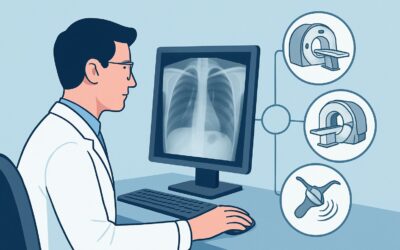
Should You Order a Routine Preoperative Chest X-ray for Asymptomatic Patients?

A 52-year-old patient is in your clinic for a preoperative evaluation before an elective inguinal hernia repair next week. They feel well, with no cough, shortness of breath, or chest pain. Their medical history is notable only for well-controlled hypertension, and they have never smoked or undergone chest surgery. As you complete the preoperative checklist, you consider whether to order a “routine” chest X-ray. Is this a necessary step to ensure anesthetic safety, or is it an outdated practice with low diagnostic yield? This article provides a detailed clinical workflow for this exact scenario: routine preoperative chest imaging for noncardiothoracic surgery in a patient with no history of chronic cardiopulmonary disease. Based on the American College of Radiology (ACR) Appropriateness Criteria, for this specific presentation, a chest radiograph is rated Usually Not Appropriate.
Who Fits This Clinical Scenario?
This guidance is narrowly focused on a specific patient population to ensure its recommendations are applied correctly. This workflow applies to patients undergoing initial evaluation for preoperative chest imaging who meet all of the following criteria:
- Procedure: The planned surgery is noncardiothoracic (e.g., orthopedic, general, urologic, gynecologic).
- Clinical Status: The patient is asymptomatic from a cardiopulmonary standpoint. There are no new or worsening symptoms like cough, dyspnea, or chest pain.
- Past Medical History: There is no known history of chronic cardiopulmonary disease (such as Chronic Obstructive Pulmonary Disease (COPD), congestive heart failure, interstitial lung disease) or prior cardiothoracic surgery.
It is critical to distinguish this scenario from similar, but distinct, clinical situations where imaging might be warranted. This guidance does not apply if:
- The patient has known cardiopulmonary disease: A patient with a history of COPD or heart failure, even if clinically stable, falls under a different ACR variant. Preoperative imaging in this group may be necessary to establish a new baseline or assess for acute exacerbations.
- The patient has new symptoms: If the preoperative history and physical exam uncover a new cough, fever, or exertional dyspnea, the patient is no longer “asymptomatic.” The imaging workup should be guided by these specific symptoms, not by a routine preoperative protocol.
- The patient is being admitted to the hospital for non-surgical reasons: Routine imaging for a general hospital admission without a specific cardiopulmonary concern is a separate clinical question.
What Diagnoses Are You Working Up in This Scenario?
When a routine preoperative chest X-ray is considered, the implicit goal is to screen for occult conditions that could increase perioperative risk. However, in an asymptomatic patient without relevant history, the pre-test probability of finding such a condition is exceedingly low. The theoretical targets of such screening include:
Occult Malignancy: The primary concern is often an unsuspected lung cancer. In an asymptomatic patient with no significant smoking history, the yield of a chest radiograph for detecting an early-stage, clinically significant lung cancer that would alter the plan for an unrelated surgery is very low.
Active Infectious Disease: Screening for active tuberculosis or other forms of pneumonia is another theoretical goal. Without symptoms like fever, cough, or weight loss, or specific epidemiologic risk factors, the likelihood of discovering active, unsuspected infection is minimal.
Cardiomegaly or Pulmonary Edema: A chest X-ray can reveal an enlarged cardiac silhouette or signs of subclinical heart failure. However, in a patient with no signs or symptoms of heart failure (e.g., no dyspnea on exertion, orthopnea, or peripheral edema), a normal physical exam is a reliable indicator of compensated status. An abnormal finding on an X-ray in this context is more likely to be a false positive or a chronic, stable finding than an acute issue requiring intervention before elective surgery.
The core issue is that the low prevalence of these conditions in this specific patient population means that screening tests are far more likely to produce false positives or identify incidental, clinically irrelevant findings than to uncover a condition that justifiably delays or cancels the planned surgery.
Why Is Routine Preoperative Chest Radiography ‘Usually Not Appropriate’?
The ACR Appropriateness Criteria panel has determined that for an asymptomatic patient with no history of cardiopulmonary disease undergoing noncardiothoracic surgery, a chest radiograph is Usually Not Appropriate. This recommendation is based on a large body of evidence demonstrating a lack of clinical benefit.
The rationale is multi-faceted:
- Low Diagnostic Yield: Studies have consistently shown that routine preoperative chest X-rays rarely reveal unexpected findings that change anesthetic or surgical management. When abnormalities are found, they are often minor, chronic, or previously known, and do not alter the perioperative plan.
- Downstream Cascade: An abnormal or indeterminate finding on a routine X-ray (e.g., a small pulmonary nodule, borderline cardiomegaly) often triggers a cascade of further testing. This can include follow-up imaging like a Computed Tomography (CT) scan, consultations, and biopsies, leading to significant patient anxiety, increased costs, and delays in the necessary primary surgery, all without improving patient outcomes.
- Radiation and Cost: While the radiation dose from a single chest radiograph is very low (☢ <0.1 mSv), it is not zero. The principle of ALARA (As Low As Reasonably Achievable) dictates that any radiation exposure must be medically justified. Without a clear clinical indication, the risk, however small, is not balanced by a tangible benefit. The financial cost of the procedure, interpretation, and any subsequent workup also contributes to the argument against routine use.
Other, higher-dose imaging modalities are even less appropriate. A CT chest without IV contrast (adult RRL ☢☢☢ 1-10 mSv) or CT chest with IV contrast (adult RRL ☢☢☢ 1-10 mSv) are both rated Usually Not Appropriate. These studies deliver substantially more radiation and have an even higher likelihood of detecting incidental findings that complicate, rather than clarify, the preoperative picture.
What’s Next? Downstream Workflow
The recommended workflow for this scenario is to rely on a thorough history and physical examination rather than routine imaging. The decision tree is straightforward:
- If the History and Physical Are Unremarkable: The patient is cleared for their planned noncardiothoracic surgery from a pulmonary standpoint. No imaging is indicated. Proceed with the scheduled operation. This is the most common and appropriate pathway for patients who fit this scenario.
- If the History and Physical Reveal New Findings: If the patient reports a new cough, worsening shortness of breath, or the exam reveals crackles or wheezing, the patient no longer fits the “routine” or “asymptomatic” criteria. The clinical question has now changed. The next step is to initiate a focused workup based on the specific symptom (e.g., chest X-ray to evaluate for pneumonia in a patient with fever and cough). The workup now follows a diagnostic, not a screening, pathway.
- If an X-ray Is Performed Anyway and Is Abnormal: This is the most common deviation from the recommended workflow. If an incidental finding like a small pulmonary nodule is discovered, it necessitates a careful, evidence-based approach. The management will depend on the finding’s characteristics and the patient’s risk factors, often guided by Fleischner Society guidelines. This may require delaying the elective surgery to complete the diagnostic workup, underscoring the potential harm of ordering a low-yield screening test in the first place.
Pitfalls to Avoid (and When to Get Help)
Navigating preoperative clearance requires avoiding several common pitfalls specific to this scenario:
- Ordering by Habit: The most significant pitfall is ordering a chest X-ray based on institutional tradition or a “checklist mentality” rather than a specific clinical indication. Always ask, “How will this result change my management?”
- Misclassifying the Patient: Be thorough in your history-taking. A patient who admits to subtle, progressive exertional dyspnea upon direct questioning is not truly asymptomatic. Similarly, a patient with a significant smoking history may warrant different considerations, potentially aligning with lung cancer screening guidelines rather than routine preoperative imaging.
- Ignoring the Asymptomatic Status: Do not order a chest X-ray “just in case” for a high-risk surgery if the patient is truly asymptomatic with no cardiopulmonary history. The type of surgery does not change the low yield of screening in this specific population.
If the preoperative assessment uncovers any new or concerning cardiopulmonary signs or symptoms, the patient should be evaluated based on that specific problem. This may involve consultation with pulmonology or cardiology before proceeding with elective surgery.
Related ACR Topics and Tools
For a comprehensive overview of all clinical variants related to this topic, please see our parent guide. Additional GigHz tools can help you apply these principles in practice.
- For breadth across all scenarios in Routine Chest Imaging, see our parent guide: Routine Chest Imaging: ACR Appropriateness Decoded.
- ACR Appropriateness Criteria Lookup: Look up adjacent or alternative clinical scenarios.
- Imaging Protocol Library: Review standard imaging techniques if a study is ultimately indicated for diagnostic reasons.
- Radiation Dose Calculator: Discuss cumulative radiation exposure with patients when multiple studies are considered.
Frequently Asked Questions
Does the patient’s age change this recommendation against routine chest X-rays?
No, for an asymptomatic patient with no history of cardiopulmonary disease, age alone is not considered an independent indication for a routine preoperative chest X-ray according to the ACR Appropriateness Criteria. The recommendation to avoid routine imaging applies to older adults as well, provided they meet the other criteria of being asymptomatic and having no relevant medical history.
What if the patient is a long-time smoker but has no symptoms?
A significant smoking history increases a patient’s risk for both lung cancer and COPD. While this patient may not fit the strict definition of this scenario (‘no history of chronic cardiopulmonary disease’), a routine preoperative chest X-ray is still often of low yield if they are truly asymptomatic. The more appropriate consideration for such a patient may be enrollment in a dedicated lung cancer screening program with low-dose CT, which is a separate clinical pathway from preoperative clearance.
Are there specific types of noncardiothoracic surgery where a chest X-ray is more likely to be needed?
The evidence suggests that even for major surgeries (e.g., major abdominal, vascular, or orthopedic procedures), a routine chest X-ray in an asymptomatic patient without cardiopulmonary disease does not improve outcomes. The decision to image should be driven by patient-specific signs and symptoms uncovered during the preoperative evaluation, not by the type or risk level of the planned surgery itself.
What if my hospital’s preoperative checklist requires a chest X-ray for all patients?
Many institutional protocols are outdated and do not reflect current evidence-based guidelines from organizations like the ACR or the American College of Physicians. This presents an opportunity for a quality improvement initiative. Citing the ACR Appropriateness Criteria can be a powerful tool for discussing a change in institutional policy to align with modern, high-value care principles and avoid unnecessary testing.
If a previous chest X-ray from a few years ago is available, should I repeat it?
No. If a patient remains asymptomatic and has no new clinical concerns, there is no indication to repeat a chest X-ray simply because a certain amount of time has passed. Repeating the imaging would be considered a routine screening test, which is rated ‘Usually Not Appropriate’ in this scenario.
Reviewed by Pouyan Golshani, MD, Interventional Radiologist — June 27, 2026