Which Imaging Is Best for Routine Follow-Up of Stable Diffuse Lung Disease?
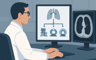
A 68-year-old patient with known idiopathic pulmonary fibrosis (IPF) returns to your clinic for a scheduled 12-month follow-up. He reports his non-productive cough is stable and his exertional dyspnea has not significantly changed since starting antifibrotic therapy. His pulmonary function tests are reassuringly stable. You now face a common clinical question: what is the most appropriate imaging study to assess for subtle disease progression, stability, or treatment response over the past year? This article provides a detailed workflow for this specific scenario—confirmed diffuse lung disease without acute deterioration. For this presentation, the American College of Radiology (ACR) rates a CT chest without IV contrast as Usually Appropriate, providing the necessary detail without unnecessary contrast or radiation.
Who Fits This Clinical Scenario?
This guidance applies to a specific and common patient population: individuals with an established diagnosis of a diffuse lung disease (DLD), such as idiopathic pulmonary fibrosis (IPF), nonspecific interstitial pneumonia (NSIP), or chronic hypersensitivity pneumonitis. The key qualifier is clinical stability. The patient is not experiencing an acute exacerbation; there is no new or worsening dyspnea, fever, cough, or hypoxemia that would suggest an infection, pulmonary embolism, or rapid fibrotic progression. The imaging is being ordered for routine, scheduled monitoring to guide long-term management.
This workflow is NOT for:
- Patients with suspected but undiagnosed DLD: These patients fall under the Initial Imaging for Suspected Diffuse Lung Disease scenario, which has a different set of diagnostic priorities and often requires high-resolution CT techniques.
- Patients with confirmed DLD and acute deterioration: A sudden decline in respiratory status requires an urgent workup to rule out superimposed processes like pneumonia, heart failure, or pulmonary embolism. This represents a distinct clinical scenario where contrast-enhanced CT may be necessary.
Applying this follow-up protocol to an acutely ill patient could delay diagnosis of a critical, treatable condition.
What Are You Assessing in This Follow-Up Scenario?
In routine follow-up of stable DLD, the imaging goal shifts from initial diagnosis to longitudinal assessment. You are not trying to identify a new disease but rather to characterize the behavior of the known disease. The key clinical questions that imaging helps answer are focused on disease trajectory and the development of complications.
Disease Progression vs. Stability: The primary goal is to determine if the fibrotic process is advancing. The radiologist will meticulously compare the new scan to prior studies, looking for subtle increases in the extent of reticulation, traction bronchiectasis, and honeycombing. The absence of change signifies stability, which is often the goal of therapy. Identifying slow, insidious progression can be the first sign that a change in medical management is warranted.
Response to Therapy: For inflammatory-mediated interstitial lung diseases (ILDs), such as those associated with connective tissue disease or hypersensitivity pneumonitis, follow-up imaging is crucial for assessing the response to immunosuppressive or antifibrotic therapy. A reduction in ground-glass opacities can indicate a positive treatment effect, whereas the development of new fibrosis might suggest treatment failure.
Detection of Complications: Patients with chronic DLD, particularly IPF, are at a significantly increased risk of developing lung cancer. Routine follow-up CT provides surveillance for new nodules or masses that could represent an early malignancy. It can also identify other complications like progressive pulmonary hypertension, evidenced by an increasing pulmonary artery diameter.
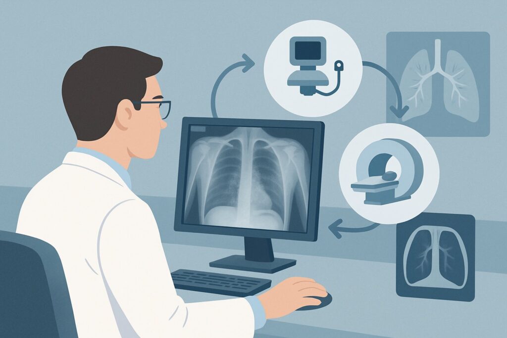
Why Is CT Chest Without IV Contrast the Recommended Study for This Presentation?
The ACR designates CT chest without IV contrast as Usually Appropriate for routine follow-up of stable DLD because it optimally balances diagnostic yield with patient safety. This modality provides exquisite detail of the lung parenchyma, which is essential for tracking the subtle changes characteristic of these diseases.
The high spatial resolution of non-contrast CT allows for clear visualization and quantification of key findings like fibrosis, ground-glass opacity, honeycombing, and emphysema. Intravenous contrast adds no significant information for assessing these parenchymal patterns and is therefore omitted to avoid the risks of contrast-induced nephropathy and allergic-like reactions. Furthermore, for patients requiring lifelong surveillance, minimizing exposure to both radiation and contrast agents is a key principle of responsible stewardship.
Let’s examine why other modalities are rated lower for this specific task:
- Radiography chest: Rated May be appropriate (Disagreement), this modality’s utility is debated. While it offers a very low radiation dose (☢ <0.1 mSv), its sensitivity for detecting subtle progression of interstitial disease is poor. A chest X-ray may appear stable even when a CT would show clear evidence of worsening fibrosis. The “Disagreement” among the ACR panel highlights that while some clinicians may use it for a quick check, it is not a reliable substitute for CT in making crucial management decisions.
- CT chest with IV contrast: Rated May be appropriate, this study is reserved for situations where there is a specific, superimposed question that requires vascular assessment. For example, if you suspect a pulmonary embolism or are evaluating a new mediastinal mass, contrast would be necessary. For routine parenchymal follow-up, however, it adds risk without benefit.
- MRI chest: Rated Usually not appropriate, MRI has limited utility for evaluating the lung parenchyma due to motion artifacts from breathing and cardiac motion, as well as lower spatial resolution compared to CT. It cannot adequately characterize the fine architectural distortion of fibrotic lung disease.
The radiation dose for a standard non-contrast chest CT is moderate (☢☢☢ 1-10 mSv). It is critical to communicate the indication for “follow-up” or “surveillance” on the order, as this often prompts the radiology department to use a low-dose or limited-scan protocol, further reducing the patient’s cumulative radiation exposure. Once you’ve decided on the top procedure, our protocol guide covers the technique and reading principles in depth: CT Chest Without Contrast.
What’s Next After CT Chest Without Contrast? Downstream Workflow
The radiologist’s comparison to prior studies is the most critical component of the report. Your subsequent actions will be guided by these findings, in conjunction with the patient’s symptoms and pulmonary function tests.
- If the CT shows stability: This is an excellent prognostic sign. It suggests the current management plan is effective at halting or significantly slowing disease progression. The typical next step is to continue the current therapy and schedule the next clinical and imaging follow-up, often in 12 to 24 months, depending on the specific disease and its known natural history.
- If the CT shows slow progression: When the report describes a subtle but definite increase in fibrotic changes (e.g., new honeycombing or increased extent of reticulation), this is a critical finding. It may prompt a multidisciplinary discussion and consideration of initiating or escalating therapy, such as starting an antifibrotic agent in a patient with progressive fibrosing ILD.
- If the CT shows a new, concerning finding: The discovery of a new solid or subsolid pulmonary nodule, for instance, shifts the workflow entirely. This triggers a separate diagnostic pathway, often guided by Fleischner Society or Lung-RADS criteria, which may involve a shorter interval follow-up CT, a PET/CT scan, or even a biopsy to rule out malignancy.
- If the CT shows improvement: In cases of inflammatory DLD treated with immunosuppression, a decrease in ground-glass opacities or consolidative change is a positive sign of treatment response. This would support continuing or tapering the current regimen.
Pitfalls to Avoid (and When to Get Help)
Navigating follow-up for DLD requires careful attention to clinical context to avoid common errors. First, do not apply this routine follow-up protocol to a patient with acute worsening; this constitutes a different clinical scenario requiring a more urgent and potentially broader workup. Second, avoid substituting a chest radiograph for a CT when a precise assessment of progression is needed to make a therapeutic decision; its low sensitivity can provide false reassurance. A third pitfall is failing to ensure low-dose techniques are used for serial scans, leading to unnecessarily high cumulative radiation exposure over a patient’s lifetime. Finally, always ensure the radiologist has access to multiple prior exams for comparison, as assessing the rate of change is often more important than the findings on a single scan. If the findings are complex or the trajectory is unclear, escalate to a multidisciplinary discussion with pulmonology and thoracic radiology.
Related ACR Topics and Tools
This article covers one specific scenario in depth. For a broader view of imaging across all clinical presentations of diffuse lung disease, from initial suspicion to acute exacerbation, please consult our comprehensive parent guide. For help with adjacent scenarios or technical details, the following GigHz resources are available.
- For breadth across all scenarios in Diffuse Lung Disease, see our parent guide: Diffuse Lung Disease: ACR Appropriateness Decoded
- To explore other clinical situations, use the ACR Appropriateness Criteria Lookup
- For technical specifications on hundreds of studies, see the Imaging Protocol Library
- To discuss cumulative exposure with patients, use the Radiation Dose Calculator
Frequently Asked Questions
Why not just get a chest X-ray for routine DLD follow-up?
A chest X-ray has very low sensitivity for the subtle changes of early disease progression. Fibrosis can worsen significantly on a CT scan while the chest X-ray appears stable. The ACR panel noted disagreement on its appropriateness, reflecting its unreliability for making key management decisions, though it may be used for a quick interval check in some contexts.
In what situation would I add IV contrast for a follow-up DLD scan?
You should add IV contrast only when you have a specific, superimposed clinical question that requires vascular evaluation. Common reasons include suspicion of pulmonary embolism in a patient with acute worsening, concern for an aortic abnormality, or characterization of a newly discovered mediastinal mass or suspicious lung nodule. For assessing the lung parenchyma alone, contrast is unnecessary.
How often should routine follow-up CT scans be performed for stable DLD?
The optimal interval is not standardized and depends on the specific type of diffuse lung disease, its rate of progression, the treatments being used, and institutional protocols. For a stable patient, intervals of 12 to 24 months are common. More rapidly progressive diseases or patients on new therapies may require more frequent imaging, such as at 6 months.
Is an MRI ever useful for following diffuse lung disease?
According to the ACR, MRI is ‘Usually not appropriate’ for this indication. The lung parenchyma is difficult to image with MRI due to low proton density and susceptibility artifacts from air-tissue interfaces. More importantly, motion from breathing and heartbeat severely degrades image quality, making it inferior to CT for visualizing the fine details of interstitial fibrosis.
What does the ACR rating ‘May be appropriate (Disagreement)’ for chest radiography mean?
This special designation means that during the ACR expert panel’s voting process, there was significant statistical variance in the ratings assigned by the panelists. It indicates a lack of consensus on the utility of the test for this specific scenario. In practice, it suggests that while some experts may find a role for it, many others do not, and its value is likely highly dependent on specific clinical factors or local practice patterns.
Reviewed by Pouyan Golshani, MD, Interventional Radiologist — May 29, 2026