What Is the Next Imaging Step for Immunocompromised Patients with a Normal Chest Radiograph?
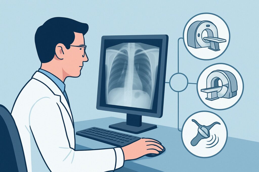
An oncology patient, three weeks post-chemotherapy for acute myeloid leukemia, develops a new fever and a dry, nonproductive cough. They are neutropenic and clinically unwell. A portable chest radiograph is obtained and read as “unremarkable,” with no clear infiltrate, effusion, or edema. The clinical team remains highly concerned for an occult pulmonary process, as the radiograph does not match the patient’s symptoms. This gap between clinical suspicion and initial imaging findings presents a critical diagnostic challenge: what is the next, most appropriate imaging study? For this specific scenario, the American College of Radiology (ACR) designates a non-contrast chest CT as Usually Appropriate, providing the necessary detail to guide life-saving therapy when a plain film falls short.
Who Fits This Clinical Scenario for Acute Respiratory Illness?
This guidance is specifically for immunocompromised patients presenting with acute respiratory signs and symptoms—such as fever, cough, or dyspnea—whose initial chest radiograph is normal, equivocal, or shows only nonspecific findings. The “immunocompromised” category is broad and includes individuals with neutropenia from chemotherapy, hematopoietic stem cell or solid organ transplant recipients, patients with advanced HIV/AIDS, or those on long-term high-dose corticosteroids or other potent immunosuppressive agents.
The key inclusion criterion is the discordance between the patient’s clinical state and their chest radiograph. The radiograph fails to provide a clear diagnosis, leaving the clinician without a definitive answer.
This workflow is distinct from several related scenarios:
- Patients needing initial imaging: If no chest radiograph has been performed yet, the decision process starts with the initial imaging variant, where a chest radiograph is the first step.
- Patients with a clearly abnormal radiograph: If the radiograph shows obvious multifocal or diffuse opacities, the workup proceeds down a different diagnostic pathway focused on characterizing a known abnormality.
- Patients with a primary suspicion of non-infectious disease: If the leading diagnosis based on clinical context is drug toxicity, pulmonary hemorrhage, or organizing pneumonia, the imaging considerations may shift.
This article focuses exclusively on the patient whose radiograph is unhelpfully normal or vague, demanding a more sensitive next step.
What Diagnoses Are You Working Up When the Chest Radiograph Is Normal?
In this vulnerable population, a normal chest radiograph can be falsely reassuring. The differential diagnosis remains broad and includes opportunistic infections and other processes that are often subtle or invisible on early plain films. The goal of advanced imaging is to detect these radiographically occult pathologies.
A primary concern is Pneumocystis jirovecii pneumonia (PJP), formerly known as PCP. This fungal infection classically presents with diffuse, bilateral ground-glass opacities. In the early stages of disease, or in patients with less severe immunosuppression, these changes can be extremely subtle and easily missed on a portable radiograph, yet they are readily apparent on a high-resolution CT scan.
Another critical consideration is invasive fungal pneumonia, most commonly caused by Aspergillus species. The earliest signs on CT are often small nodules or areas of consolidation, which may be surrounded by a “halo” of ground-glass opacity representing hemorrhage. These findings are highly suggestive of the diagnosis but are typically far too small to be resolved on a chest X-ray.
Viral pneumonias, such as those caused by Cytomegalovirus (CMV), respiratory syncytial virus (RSV), or influenza, can also manifest with subtle ground-glass changes, centrilobular nodules, or a tree-in-bud pattern. These findings indicate inflammation in the small airways and surrounding lung tissue, patterns that are hallmarks of CT imaging but are generally below the detection threshold of radiography.
Finally, early bacterial pneumonias or infections with atypical organisms like Nocardia can begin as small, ill-defined nodules or patchy consolidations that are not yet dense enough to be visible on a radiograph. CT can identify these early-stage infections, allowing for prompt initiation of targeted antimicrobial therapy.
Why Is a Non-Contrast Chest CT the Recommended Study for This Presentation?
The ACR rates CT chest without IV contrast as Usually Appropriate because it directly and effectively addresses the key diagnostic questions in this scenario while minimizing patient risk. The superior contrast and spatial resolution of CT compared to radiography make it highly sensitive for detecting the subtle parenchymal abnormalities—ground-glass opacities, micronodules, and early consolidation—that characterize opportunistic infections.
The decision to omit intravenous contrast is deliberate. The primary diagnostic targets are within the lung parenchyma and airways. IV contrast does not enhance these findings and is therefore unnecessary for the initial evaluation. Forgoing contrast avoids the risks of contrast-induced nephropathy in patients who may already have renal compromise from their underlying disease or medications. It also eliminates the risk of allergic-like reactions and streamlines the imaging process, allowing for faster acquisition in a potentially unstable patient.
Alternatives are rated lower for specific reasons:
- CT chest with IV contrast is rated May be appropriate. While it provides the same excellent parenchymal detail, it adds the risks associated with contrast media without offering significant diagnostic benefit for the primary question. It should be reserved for cases where there is a specific secondary concern, such as a suspected pulmonary embolism or a complex abscess requiring vascular assessment.
- MRI chest without or with IV contrast is rated Usually not appropriate. MRI is significantly limited by motion artifact from breathing and cardiac motion, and it has inferior spatial resolution for evaluating the fine architecture of the lung parenchyma. It is not the correct tool for identifying the subtle ground-glass changes or small nodules characteristic of early opportunistic infections.
The recommended non-contrast CT involves a relative radiation level of ☢☢☢ (1-10 mSv) for adults, a dose that is justified by the high diagnostic yield and the critical importance of reaching a timely diagnosis in this high-risk population. Once you’ve decided on CT chest without IV contrast, our protocol guide covers the technique, dose considerations, and reading principles: CT Chest Without Contrast.
What’s Next After CT chest without IV contrast? Downstream Workflow
The results of the non-contrast chest CT will guide the subsequent diagnostic and therapeutic pathway. The goal is to move from imaging findings to a specific etiologic diagnosis.
If the CT demonstrates diffuse, bilateral ground-glass opacities, the primary suspicion shifts strongly toward PJP or a viral process. The immediate next step is typically bronchoscopy with bronchoalveolar lavage (BAL). The BAL fluid can be sent for specific stains (for PJP), viral PCR panels, and other microbiologic cultures. Empiric treatment for PJP is often started immediately after the CT, pending BAL results.
If the CT reveals one or more nodules, particularly with a halo sign or air-crescent sign, invasive fungal pneumonia becomes the leading diagnosis. In addition to BAL, serum biomarkers like galactomannan and beta-D-glucan are crucial. Antifungal therapy should be initiated promptly, as these infections can progress rapidly. A percutaneous biopsy may be considered for a solitary, accessible lesion if less invasive methods are non-diagnostic.
If the CT is negative and shows no parenchymal abnormality, it provides strong evidence against a significant pulmonary infection. The clinical team should then pivot to investigate non-pulmonary sources of fever and consider alternative diagnoses such as drug fever, line infection, or sinusitis.
If the CT findings are indeterminate or nonspecific (e.g., minor atelectasis, scattered bronchial wall thickening), the results must be correlated closely with the clinical picture. If clinical suspicion remains high, proceeding to BAL may still be warranted to sample the distal airways, even without a clear parenchymal target.
Pitfalls to Avoid (and When to Get Help)
Navigating this scenario requires careful attention to clinical context to avoid common errors.
- Delaying the CT: Do not be overly reassured by a normal chest radiograph in a symptomatic, high-risk patient. A delay in diagnosis can lead to significant morbidity and mortality.
- Ordering the wrong study: Ordering a contrast-enhanced CT by default is a common pitfall. For this indication, non-contrast is sufficient and safer. Conversely, if pulmonary embolism is a genuine, primary concern, a dedicated CT pulmonary angiogram (CTPA) protocol is required, not a standard contrast-enhanced chest CT.
- Ignoring pediatric dose: For pediatric patients, the radiation dose for a chest CT is a more significant consideration (ACR pediatric RRL ☢☢☢☢, 3-10 mSv). Ensure that low-dose pediatric protocols are used.
- Misinterpreting subtle findings: Subtle ground-glass opacity can be mistaken for motion artifact or poor inspiration. Close collaboration with the interpreting radiologist is key to ensuring these findings are given appropriate clinical weight.
If the patient is rapidly deteriorating or hemodynamically unstable, escalate immediately to critical care specialists and consider an urgent, portable CT if available and clinically appropriate.
Related ACR Topics and Tools
This article covers one specific clinical variant. For a comprehensive overview of all scenarios, from initial imaging to follow-up, please consult our parent guide. For tools to help select the right study and understand the details, see the resources below.
- Parent Topic Hub: For breadth across all scenarios in Acute Respiratory Illness in Immunocompromised Patients, see our parent guide: Acute Respiratory Illness in Immunocompromised Patients: ACR Appropriateness Decoded.
- ACR Appropriateness Criteria Lookup: Imaging Appropriateness Selector — for adjacent scenarios
- Imaging Protocol Library: Imaging Protocol Library — for technique on the recommended study
- Radiation Dose Calculator: Radiation Dose Calculator — for cumulative dose conversations
Frequently Asked Questions
Why not just repeat the chest radiograph in 24-48 hours instead of getting a CT?
While a repeat radiograph might eventually show an evolving process, it lacks the sensitivity to detect early-stage opportunistic infections. In a high-risk, immunocompromised patient, the delay in diagnosis could lead to clinical deterioration and worse outcomes. A CT provides a definitive answer much earlier in the disease course.
Is a low-dose chest CT sufficient for this indication?
Yes, in many cases, a low-dose or ultra-low-dose non-contrast chest CT protocol can provide sufficient diagnostic information for detecting parenchymal abnormalities like ground-glass opacities and nodules. This approach helps minimize radiation exposure, which is an important consideration in patients who may require frequent imaging. Consult with your radiology department about their specific low-dose protocols.
If the CT is negative, can I confidently rule out a pulmonary infection?
A negative high-resolution non-contrast chest CT has a very high negative predictive value for significant parenchymal lung disease. While it cannot rule out tracheobronchitis or a very early infection, it makes a clinically significant pneumonia highly unlikely. If symptoms persist, the focus should shift to non-pulmonary sources of infection or non-infectious causes.
Should I order a CT with contrast if the patient also has pleuritic chest pain?
If the primary concern driving the chest pain is pulmonary embolism (PE), then a dedicated CT pulmonary angiogram (CTPA) is the appropriate study, not a standard contrast-enhanced chest CT. If PE is not suspected, a non-contrast CT is still the best initial test, as it can identify peripheral parenchymal processes that may cause pleuritic pain.
What if the patient is too unstable to transport to the CT scanner?
In a critically ill patient who cannot be safely transported, the clinical team must weigh the risks and benefits. Options may include bedside bronchoscopy with BAL without prior imaging or stabilizing the patient in the ICU before attempting transport. Some institutions have portable CT scanners for ICU use, which can be an invaluable tool in this situation.
Reviewed by Pouyan Golshani, MD, Interventional Radiologist — May 26, 2026