What Is the Next Step for an Incidental Pulmonary Nodule on Chest X-Ray?

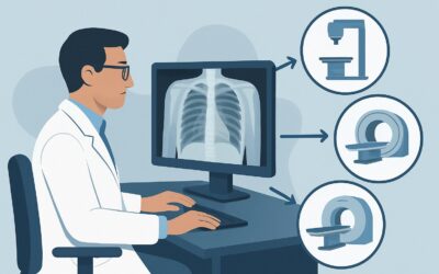
It’s late in the afternoon clinic, and you’re reviewing pre-operative labs and imaging for a 58-year-old patient scheduled for a cholecystectomy. The routine chest radiograph, ordered for pre-anesthesia clearance, is unremarkable except for a single, well-circumscribed 9 mm opacity in the right mid-lung that was not present five years ago. The patient is asymptomatic. This finding raises an immediate clinical question: what is the appropriate next imaging study to evaluate this incidentally detected, indeterminate pulmonary nodule? This article provides a focused, evidence-based workflow for this exact scenario. According to the American College of Radiology (ACR) Appropriateness Criteria, the definitive next step is a CT chest without IV contrast, which is rated Usually Appropriate.
## Who Fits This Clinical Scenario?
This guidance is specifically for an adult patient, 35 years of age or older, who has an indeterminate pulmonary nodule detected incidentally on a chest radiograph (X-ray).
The key elements defining this scenario are:
- Patient Age: The patient is 35 or older. Malignancy risk increases with age, altering the pre-test probability and justifying a more definitive workup than in a younger patient.
- Incidental Finding: The nodule was discovered on an imaging study performed for an unrelated reason. The patient has no signs or symptoms concerning for lung cancer, such as hemoptysis, unexplained weight loss, or new cough.
- Initial Modality: The nodule was first seen on a two-dimensional chest radiograph. This is a critical distinction, as the workup for a nodule initially detected on a CT scan follows a different pathway (e.g., Fleischner Society guidelines).
- Indeterminate Nature: The nodule lacks definitively benign features on the radiograph, such as central, diffuse, or popcorn-like calcification. It is simply an opacity that requires further characterization.
This workflow does not apply to patients with a known history of cancer, as a new pulmonary nodule in this context raises immediate concern for metastasis and often requires a different imaging approach (e.g., PET/CT or contrast-enhanced CT). It also does not apply to patients who are symptomatic or have a clinical suspicion of active infection.
## What Diagnoses Are You Working Up in This Scenario?
When an indeterminate pulmonary nodule is found on a chest radiograph, the primary goal of the subsequent workup is to differentiate between malignant and benign causes. The differential diagnosis is broad, but the following are the most critical considerations.
The most consequential possibility is a primary lung malignancy, such as adenocarcinoma. This is the diagnosis that must be excluded. The likelihood depends heavily on patient risk factors, including smoking history, family history of lung cancer, and occupational exposures. CT imaging is essential for evaluating nodule characteristics associated with malignancy, such as size, spiculated margins, and growth over time.
A very common benign cause is an infectious granuloma. These are organized collections of immune cells that form in response to infections, most commonly from fungal sources (like histoplasmosis, prevalent in certain geographic regions) or mycobacteria (like previous tuberculosis). On CT, granulomas often have features like dense, central calcification that confirm their benign nature, effectively ending the workup.
Less common but important benign entities include benign neoplasms like a pulmonary hamartoma. These tumors are composed of a mix of cartilage, fat, and other tissues. The presence of fat density or “popcorn” calcification on a non-contrast CT is pathognomonic and requires no further follow-up.
Finally, an intrapulmonary lymph node can appear as a small, indeterminate nodule on a radiograph. These are typically benign, well-defined, and located in the lower lobes near the pleura. CT can often confirm their characteristic location and appearance, resolving the initial uncertainty.
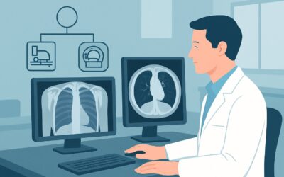
## Why Is CT Chest Without IV Contrast the Recommended Study for This Presentation?
The ACR rates CT chest without IV contrast as Usually Appropriate because it directly and efficiently answers the primary clinical question: what are the precise characteristics of the nodule seen on the radiograph? It provides the necessary anatomical detail to stratify risk and determine the next steps.
A non-contrast CT excels in this scenario for several key reasons:
- Superior Characterization: CT offers vastly superior spatial and contrast resolution compared to a chest radiograph. It can precisely measure the nodule’s size, define its borders (smooth vs. spiculated), assess its density, and, most importantly, detect specific features of benignity like fat or particular patterns of calcification that are often invisible on X-ray. It also definitively confirms the finding is a true intrapulmonary nodule and not a summation shadow from overlapping structures.
- No Added Value from Contrast: For the initial characterization of an indeterminate nodule, intravenous contrast is not necessary. The key diagnostic information—size, morphology, density, and calcification—is fully assessed on non-contrast images. Administering IV contrast adds potential risks (e.g., allergic reaction, contrast-induced nephropathy) and cost without improving the primary diagnostic yield for this indication.
- Appropriate Radiation Dose: While a CT involves more radiation than a radiograph, a modern non-contrast chest CT is a moderate-dose study (ACR Relative Radiation Level ☢☢☢, 1-10 mSv). This exposure is justified to resolve the ambiguity of a potential malignancy and prevent the delay in diagnosis that could result from less definitive follow-up.
### Why Other Studies Are Less Appropriate
- Radiography chest: A follow-up chest X-ray is rated May be appropriate. While it involves a very low radiation dose (☢, <0.1 mSv), it provides limited new information. It can assess for short-term growth, but it cannot characterize the nodule's internal features. Relying on serial radiographs can lead to significant delays in diagnosing a malignancy.
- CT chest with IV contrast: This is rated Usually not appropriate. As noted, contrast does not aid in the initial characterization and should be reserved for specific indications later in a workup, such as assessing for mediastinal invasion or hilar adenopathy if a nodule is deemed highly suspicious.
- FDG-PET/CT: This is also rated Usually not appropriate as a next step. PET/CT is a functional imaging study used for staging known cancer or evaluating nodules that remain indeterminate after a full diagnostic CT workup. It has a high radiation dose (☢☢☢☢, 10-30 mSv) and is not the correct tool for initial characterization.
Once you’ve decided on the recommended study, ensuring proper execution is key. For a detailed overview of the imaging technique, reading principles, and dictation, see our comprehensive protocol guide: CT Chest Without Contrast.
## What’s Next After CT Chest Without IV Contrast? Downstream Workflow
The results of the non-contrast chest CT will guide the subsequent management, which typically transitions to risk-based follow-up protocols like those from the Fleischner Society.
- If the CT confirms a definitively benign finding: If the nodule demonstrates features like fat (suggesting a hamartoma) or a benign pattern of calcification (central, laminated, diffuse, or popcorn), the workup is complete. No further imaging is needed. Similarly, if the CT reveals the “nodule” was actually a summation shadow, rib artifact, or other non-pulmonary finding, the workup ends.
- If the CT confirms an indeterminate solid nodule: Management now depends on the nodule’s size and the patient’s clinical risk for lung cancer (e.g., smoking history). This is where the workflow moves into one of the sibling ACR scenarios. For example:
- If the nodule measures less than 6 mm, follow-up may not be required in a low-risk patient, or a single follow-up CT at 12 months may be considered for a high-risk patient.
- If the nodule measures 6 mm or greater, serial CT surveillance is typically recommended, with the interval and duration determined by the initial nodule size and patient risk factors.
- If the CT shows a subsolid nodule (part-solid or ground-glass): These have a different natural history and require a distinct follow-up pathway, often with longer surveillance intervals, as they may represent slower-growing adenocarcinomas.
- If the CT reveals highly suspicious features: If the nodule is large, has spiculated margins, or is associated with lymphadenopathy, the workflow accelerates. The next step is no longer surveillance but rather pursuing a definitive diagnosis via biopsy (image-guided or bronchoscopic) and/or a PET/CT for staging.
## Pitfalls to Avoid (and When to Get Help)
Navigating this common clinical scenario requires careful attention to detail to avoid several potential missteps.
1. Applying this workflow to a symptomatic patient: If a patient has symptoms like hemoptysis, chest pain, or unexplained weight loss, they do not have an “incidental” nodule. This is a cancer workup from the start and requires a more aggressive diagnostic approach.
2. Ordering a contrast-enhanced CT by default: Many institutional CT protocols default to including IV contrast. For this specific indication, you must explicitly order a “CT chest without IV contrast” to avoid unnecessary risk, cost, and potential artifacts.
3. Failing to close the loop on follow-up: The single biggest pitfall is identifying a nodule and not ensuring the recommended CT is performed and the results are reviewed and acted upon. Robust tracking systems are critical for patient safety.
If the non-contrast CT reveals a large, highly suspicious mass or extensive mediastinal and hilar lymphadenopathy, the situation has escalated beyond a simple nodule workup. This warrants an urgent referral to a pulmonologist or thoracic oncologist for expedited tissue diagnosis and staging.
## Related ACR Topics and Tools
This article covers one specific variant within the broader topic of indeterminate pulmonary nodules. For a comprehensive overview of all related clinical scenarios, from nodules found on CT to management in younger patients, please see our parent guide.
- For breadth across all scenarios in Incidentally Detected Indeterminate Pulmonary Nodule, see our parent guide: Incidentally Detected Indeterminate Pulmonary Nodule: ACR Appropriateness Decoded
To explore other scenarios, imaging techniques, or radiation dose considerations, these GigHz tools can help:
- ACR Appropriateness Criteria Lookup — for adjacent scenarios
- Imaging Protocol Library — for technique on the recommended study
- Radiation Dose Calculator — for cumulative dose conversations
Frequently Asked Questions
Why not just get a follow-up chest X-ray in a few months to check for growth?
While a follow-up chest X-ray is rated ‘May be appropriate’ by the ACR, it is a less ideal strategy. It can only assess for significant interval growth and provides no information about the nodule’s internal characteristics (like calcification or fat) that could immediately confirm it as benign. Relying on a chest X-ray can delay the diagnosis of a potentially curable early-stage lung cancer. A non-contrast CT is the definitive next step to fully characterize the nodule and stratify risk.
Is IV contrast ever needed for a pulmonary nodule workup?
Yes, but not for the initial characterization of an indeterminate nodule found on a radiograph. IV contrast is used later in the workup if a nodule is found to be highly suspicious for malignancy. In that context, a contrast-enhanced CT or PET/CT helps evaluate for mediastinal or hilar lymph node involvement and assess for potential invasion of adjacent vascular structures, which is critical for staging and surgical planning.
What if the patient is under 35 years old?
This specific ACR guidance applies to adults aged 35 and older. In younger patients, the pre-test probability of malignancy is significantly lower, and the lifetime attributable risk from radiation is higher. While the ACR does not have a separate variant for this exact scenario in younger adults, clinical practice often favors a more conservative approach, such as a short-interval follow-up chest radiograph, especially for smaller nodules. A CT may still be warranted if the nodule is large or has suspicious features on the initial X-ray.
The radiologist’s report on the X-ray is vague. How do I know if the nodule is truly ‘indeterminate’?
A nodule is considered indeterminate on a chest radiograph if it lacks classic benign features. A definitively benign nodule would show specific calcification patterns (dense, central, laminated, or ‘popcorn’) or be demonstrably stable for at least two years on prior imaging. If the report describes a ‘non-calcified nodule,’ ‘focal opacity,’ or ‘soft tissue density’ without these benign signs, it should be treated as indeterminate and warrants further evaluation with a non-contrast chest CT.
Does the patient’s smoking history change the recommendation for a non-contrast CT?
No, the recommendation to obtain a non-contrast chest CT as the next step remains the same regardless of smoking history. However, the patient’s risk factors, particularly smoking history, become critically important for interpreting the CT results and determining the downstream management (e.g., the intensity and duration of CT surveillance) according to guidelines like those from the Fleischner Society.
Reviewed by Pouyan Golshani, MD, Interventional Radiologist — May 30, 2026