

Your Patient Has an SMA Embolus and Peritonitis: What’s the ACR-Guided Next Step?
ACR-guided imaging workflow for Recent onset abdominal pain, peritoneal signs, and known atrial fibrillation. CTA shows filling defect in the proximal SMA
Part of the imaging decision system doctors were never taught to navigate at speed: which study to order, which protocol, and what the published appropriateness guidance actually says for IR imaging. Plain-English references, scenario by scenario.
ACR-guided imaging workflow for Recent onset abdominal pain, peritoneal signs, and known atrial fibrillation. CTA shows filling defect in the proximal SMA
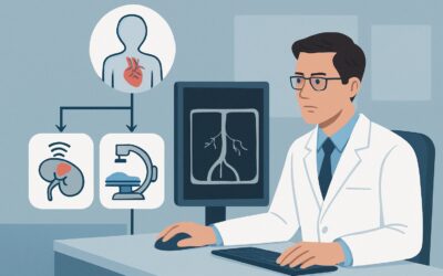
ACR-guided imaging workflow for Recent onset abdominal pain, no peritoneal signs, and known atrial fibrillation. CTA shows calcified atherosclerotic plaque

ACR-guided imaging workflow for Recent onset abdominal pain, no peritoneal signs, and known atrial fibrillation. CTA shows filling defect in proximal SMA c
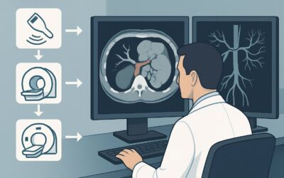
ACR-guided imaging workflow for Ductal cholangiocarcinoma: Hilar cholangiocarcinoma, greater than 3 cm with poorly defined margins, vascular invasion, and

ACR-guided imaging workflow for Intrahepatic cholangiocarcinoma: Peripheral hepatic lobar cholangiocarcinoma, less than 3 cm; no biliary ductal dilatation,

ACR-guided imaging workflow for Hepatocellular cancer: Solitary or multifocal disease with vascular invasion, cirrhotic. (Management of Liver Cancer): reco
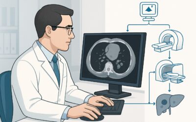
ACR-guided imaging workflow for Hepatocellular cancer: Multifocal, bilobar disease, at least 1 tumor greater than 5 cm, cirrhotic. (Management of Liver Can
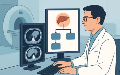
ACR-guided imaging workflow for Hepatocellular cancer: Solitary tumor 3 to 5 cm, cirrhotic. (Management of Liver Cancer): recommended study, differential,
ACR-guided imaging workflow for Lower gastrointestinal tract bleeding. Obscure (nonlocalized) recurrent bleeding in a hemodynamically stable patient (assum
ACR-guided imaging workflow for Lower gastrointestinal tract bleeding. Transcatheter arteriography localized the bleeding site and treatment was attempted.