

What Is the Next Step for Refractory Ascites and Worsening Renal Function?
ACR-guided imaging workflow for Ascites. Child-Pugh class B cirrhotic with chronic ascites undergoing weekly large-volume paracentesis; rapidly declining r
Part of the imaging decision system doctors were never taught to navigate at speed: which study to order, which protocol, and what the published appropriateness guidance actually says for IR imaging. Plain-English references, scenario by scenario.
ACR-guided imaging workflow for Ascites. Child-Pugh class B cirrhotic with chronic ascites undergoing weekly large-volume paracentesis; rapidly declining r

ACR-guided imaging workflow for Ascites. Child-Pugh class B cirrhotic with chronic ascites despite daily diuretic therapy and low-sodium diet. (Radiologic

ACR-guided imaging workflow for Ascites. Initial therapy for Child-Pugh class B cirrhotic asymptomatic patient with small-volume ascites. (Radiologic Manag

ACR-guided imaging workflow for Acute variceal bleeding. Child-Pugh class C, cirrhotic with hepatocellular carcinoma, branch portal vein tumor thrombus, an

ACR-guided imaging workflow for Acute variceal bleeding. Child-Pugh class C, cirrhotic with active esophageal and junctional variceal hemorrhage, previousl

ACR-guided imaging workflow for Acute variceal bleeding. Child-Pugh class B, cirrhotic with active esophageal variceal hemorrhage, MELD 12, previously trea

ACR-guided imaging workflow for Acute variceal bleeding. Child-Pugh class A, cirrhotic with index bleed from acute esophageal variceal hemorrhage, MELD 10,

ACR-guided imaging workflow for Acute iliofemoral DVT in a pregnant patient with moderate to severe symptoms. (Radiologic Management of Iliofemoral Venous

ACR-guided imaging workflow for Iliofemoral DVT with persistent moderate symptoms at least 3 months after initial treatment with anticoagulation alone. (Ra
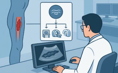
ACR-guided imaging workflow for Acute femoropopliteal DVT with mild to moderate symptoms present for less than 14 days, otherwise healthy. (Radiologic Mana