Should You Order MR Arthrography for a Suspected Hip Labral Tear with Normal Radiographs?

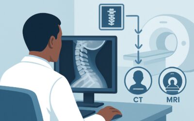
A 34-year-old marathon runner presents to your clinic with four months of insidious-onset, deep anterior hip pain. They describe a catching sensation with pivoting movements and pain that worsens after long runs. The physical exam is notable for a positive FADIR (flexion, adduction, internal rotation) test, but initial anteroposterior pelvis and lateral hip radiographs are read as normal, showing no signs of osteoarthritis, fracture, or significant bony deformity. You suspect an acetabular labral tear, but the normal radiographs leave you at a diagnostic crossroads. What is the most effective next imaging study to confirm or exclude this diagnosis and guide management? For this specific clinical scenario, the American College of Radiology (ACR) Appropriateness Criteria rate MR arthrography of the hip as Usually Appropriate.
Who Fits This Clinical Scenario for a Suspected Labral Tear?
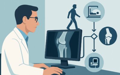
This guidance applies to a well-defined patient population: individuals with chronic hip pain where a labral tear is high on the differential diagnosis, and initial radiographs have already been obtained and are unrevealing.
Inclusion criteria for this workflow:
- Symptom Duration: The hip pain is chronic, present for weeks to months.
- Symptom Character: Pain is often located in the anterior hip or groin and may be accompanied by mechanical symptoms like clicking, catching, locking, or a feeling of instability.
- Physical Exam: Findings are suggestive of intra-articular pathology, such as a positive FADIR or FABER test.
- Prior Imaging: Anteroposterior (AP) pelvis and hip radiographs have been performed and are negative or nondiagnostic for the cause of pain (e.g., no significant osteoarthritis, dysplasia, or fracture).
This workflow is NOT for patients who:
- Present for initial imaging: If the patient has had no prior imaging, the first step is typically radiography. This is covered in the ACR variant for Chronic Hip Pain, Initial Imaging.
- Have pain localized to the greater trochanter: If symptoms and physical exam point to a lateral process like gluteal tendinopathy or trochanteric bursitis, the imaging strategy differs. This falls under the variant for suspected extra-articular abnormalities.
- Have clear bony abnormalities on radiographs: If radiographs demonstrate findings of femoroacetabular impingement (FAI) or hip dysplasia, the imaging question shifts from primary diagnosis to preoperative planning, which is a distinct clinical scenario.
What Diagnoses Are You Working Up in This Scenario?
When ordering advanced imaging for a suspected labral tear, you are evaluating a spectrum of intra-articular pathologies that are invisible on plain radiographs. The differential diagnosis guides the choice of imaging modality and protocol.
Acetabular Labral Tear This is the primary diagnosis of concern. The labrum is a fibrocartilaginous ring that deepens the acetabulum, contributing to hip stability and joint lubrication. Tears can result from acute trauma, repetitive microtrauma, or underlying bony abnormalities like FAI. They are a common source of mechanical symptoms and pain.
Femoroacetabular Impingement (FAI) FAI is a condition where abnormal contact between the femoral head-neck junction and the acetabular rim leads to damage of the labrum and articular cartilage. While radiographs may suggest FAI, they can miss subtle cam or pincer morphology. Advanced imaging provides a detailed three-dimensional assessment of the bony anatomy that is critical for diagnosis and surgical planning.
Chondral Injury or Early Osteoarthritis Damage to the articular cartilage often coexists with labral tears and FAI. Radiographs are insensitive to early cartilage loss, only showing joint space narrowing in later stages. MRI is highly sensitive for detecting chondral defects, delamination, and edema in the subchondral bone, which are important prognostic findings.
Ligamentum Teres Tear Though less common, a tear of the ligamentum teres—a ligament connecting the femoral head to the acetabulum—can cause deep, persistent hip pain and instability. This structure is best visualized with advanced imaging, particularly MR arthrography.
Why Is MR Arthrography the Top Study for a Suspected Labral Tear?
The ACR designates both MR arthrography and non-contrast MRI of the hip as Usually Appropriate for this scenario, with MR arthrography often considered the gold standard for its superior visualization of intra-articular structures.
The primary advantage of MR arthrography hip is the injection of dilute gadolinium-based contrast directly into the joint space under fluoroscopic or ultrasound guidance. This technique distends the joint capsule, outlining the labrum and articular cartilage in high relief. The contrast fills any clefts or tears in the labrum, making them significantly more conspicuous than on a non-arthrographic study. This markedly improves the sensitivity and diagnostic confidence for detecting labral tears, chondral defects, and ligamentum teres injuries.
A high-quality MRI hip without IV contrast is also rated Usually Appropriate and serves as an excellent non-invasive alternative. Modern 3-Tesla (3T) MRI scanners with optimized protocols can achieve high diagnostic accuracy for labral tears without an injection. The choice between arthrography and a non-contrast study often depends on institutional preference, radiologist expertise, and patient factors. A non-contrast MRI is often preferred as the initial advanced imaging step, with arthrography reserved for cases where the non-contrast study is equivocal or a higher degree of certainty is required.
Why are other studies rated lower?
- CT arthrography hip (May be appropriate): This study also uses intra-articular contrast and provides excellent detail of the labrum and osseous structures. However, it is less sensitive for evaluating articular cartilage and surrounding soft tissues and involves ionizing radiation (ACR RRL=☢☢☢, 1-10 mSv). It is primarily reserved for patients with contraindications to MRI.
- US hip (Usually not appropriate): While ultrasound is valuable for assessing superficial structures like tendons and bursae around the hip, it cannot penetrate deep enough to reliably visualize the acetabular labrum or other intra-articular structures. Its role in this specific scenario is therefore limited.
Both recommended MRI options are radiation-free (ACR RRL=O, 0 mSv), making them ideal for the often younger, active patient population affected by labral tears.
Once you’ve decided on the best study for your patient, our protocol guide covers the technique, contrast, and reading principles: MRI Hip Without Contrast.
What’s Next After an MRI? Downstream Workflow
The results of the MRI or MR arthrogram will directly guide the subsequent clinical pathway. The goal is to correlate the imaging findings with the patient’s specific symptoms and functional goals to create a tailored treatment plan.
- If the study is positive for a labral tear and/or FAI: The next step is typically a referral to an orthopedic surgeon specializing in hip preservation. The surgeon will discuss treatment options, which may range from conservative management (physical therapy, activity modification, injections) to surgical intervention (hip arthroscopy for labral repair and/or osteochondroplasty for FAI). The detailed anatomical information from the MRI is crucial for surgical planning.
- If the study is negative for intra-articular pathology: This is a critical branch point. The absence of a labral tear or chondral injury prompts a re-evaluation of the differential diagnosis. The focus should shift to extra-articular causes of hip and groin pain. This may include iliopsoas tendinopathy, athletic pubalgia (sports hernia), adductor strain, or referred pain from the lumbar spine. Further clinical assessment or different imaging may be required, potentially aligning with the ACR variant for suspected extra-articular abnormalities.
- If the study is indeterminate or shows unexpected findings: Equivocal findings may warrant a discussion with the interpreting radiologist. In some cases, a diagnostic/therapeutic intra-articular injection, rated May be appropriate, can be a useful next step. Injecting local anesthetic and corticosteroid into the hip joint can help confirm an intra-articular source of pain if the patient experiences significant, albeit temporary, relief.
Pitfalls to Avoid (and When to Get Help)
Navigating the workup for a suspected labral tear requires careful attention to detail to avoid common missteps.
- Pitfall 1: Skipping Radiographs. Never order an MRI for chronic hip pain without first obtaining plain radiographs. Radiographs are essential for ruling out significant osteoarthritis, dysplasia, or other bony abnormalities that would fundamentally change the diagnosis and management plan.
- Pitfall 2: Ordering a “Routine” Lumbar Spine MRI. While hip pain can be referred from the spine, ordering a lumbar MRI before thoroughly evaluating the hip joint itself can lead to delayed diagnosis and treatment of intra-articular hip pathology.
- Pitfall 3: Misinterpreting a “Normal” MRI Report. A non-contrast MRI performed on a lower-field-strength magnet (1.5T) or without a dedicated labral tear protocol may have limited sensitivity. If clinical suspicion remains high despite a negative non-contrast MRI, consider discussing the case with a musculoskeletal radiologist or proceeding to MR arthrography.
If a patient presents with constitutional symptoms (fever, weight loss), a history of malignancy, or acute-on-chronic pain with an inability to bear weight, escalate care for an urgent workup to rule out infection, tumor, or an occult fracture.
Related ACR Topics and Tools
This article focuses on one specific scenario. For a comprehensive overview of all clinical variants and imaging modalities for chronic hip pain, or to explore adjacent clinical questions, the following resources are essential.
- For breadth across all scenarios in Chronic Hip Pain, see our parent guide: Chronic Hip Pain: ACR Appropriateness Decoded.
- ACR Appropriateness Criteria Lookup: Search and compare imaging recommendations for thousands of clinical scenarios.
- Imaging Protocol Library: Access detailed, step-by-step protocols for performing the recommended studies.
- Radiation Dose Calculator: Quantify and discuss radiation exposure with patients when considering CT or other ionizing modalities.
Frequently Asked Questions
Is MR arthrography always better than a non-contrast MRI for a suspected labral tear?
Not necessarily. While MR arthrography is often considered the most sensitive test, a high-quality 3T non-contrast MRI can have comparable diagnostic accuracy. The ACR rates both as ‘Usually Appropriate.’ The choice often depends on institutional protocols, radiologist preference, and patient factors. Many centers start with a non-contrast MRI as a less invasive first step.
What if my patient is claustrophobic or has a contraindication to MRI?
If a patient cannot undergo an MRI, CT arthrography is a viable alternative and is rated ‘May be appropriate’ by the ACR. It provides excellent visualization of the labrum and bony anatomy but is less effective for evaluating cartilage and involves ionizing radiation. A thorough discussion of the risks and benefits is necessary.
Why isn’t an MRI with intravenous (IV) contrast recommended?
An MRI of the hip with and/or without IV contrast is rated ‘Usually not appropriate’ for this specific scenario. Standard IV contrast enhances vascular structures and areas of inflammation but does not distend the joint capsule or seep into labral tears effectively. Therefore, it does not significantly improve the detection of labral or chondral pathology compared to a non-contrast study and is less effective than direct intra-articular contrast (arthrography).
My patient’s radiograph report mentioned a ‘possible cam lesion.’ Does this change the workflow?
Yes, it might. If radiographs are suspicious for femoroacetabular impingement (FAI) or dysplasia, the clinical question shifts slightly. While the recommended next imaging study (MRI or MR arthrography) is often the same, the primary goal becomes characterizing the bony morphology for surgical planning in addition to diagnosing the labral tear. This aligns more closely with the ACR variant for ‘Suspect impingement or dysplasia.’
Can a diagnostic hip injection be performed at the same time as the MR arthrogram?
Yes, this is a common and efficient practice. During the MR arthrogram procedure, after confirming intra-articular needle placement but before injecting the gadolinium contrast, a small amount of local anesthetic (like lidocaine or bupivacaine) can be injected. The patient can then assess their pain relief over the next few hours. This combines the diagnostic power of the MRI with the therapeutic and prognostic information of a selective joint block.
Reviewed by Pouyan Golshani, MD, Interventional Radiologist — May 21, 2026