What’s the Best Initial Imaging for C-Spine Trauma When NEXUS Criteria Are Met?
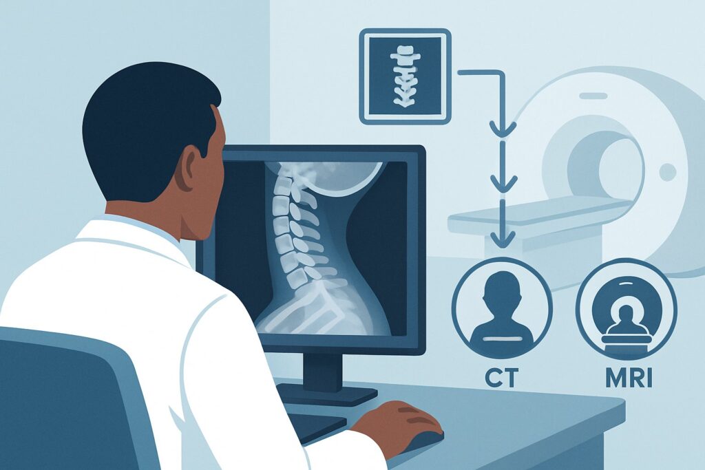
It’s 2 a.m. in the emergency department, and you’re evaluating a 24-year-old male brought in by paramedics after a motor vehicle collision. He was the restrained driver, the airbags deployed, and he’s now immobilized on a backboard with a cervical collar. He is alert, not intoxicated, and complains of midline cervical tenderness on palpation. His neurologic exam is normal. Based on the National Emergency X-Radiography Utilization Study (NEXUS) criteria, he requires imaging to rule out a cervical spine injury. The immediate question is which study to order first. This article details the American College of Radiology (ACR) Appropriateness Criteria workflow for this exact scenario: an adult with acute blunt cervical spine trauma who meets clinical criteria for imaging. For this presentation, the ACR designates **CT cervical spine without IV contrast** as *Usually Appropriate*.
Who Fits This Clinical Scenario?
This guidance applies to a specific and common patient population: individuals aged 16 years or older who have sustained acute blunt trauma and require cervical spine imaging based on validated clinical decision rules. The two most widely used rules are the NEXUS criteria and the Canadian C-Spine Rule (CCR). If a patient meets the criteria for imaging under either of these rules—for example, due to midline cervical spine tenderness, a dangerous mechanism of injury, or the inability to rotate their neck—this workflow is the appropriate starting point.
It is crucial to distinguish this scenario from others that may appear similar but require a different diagnostic approach. This guidance does not apply to:
- Patients who can be clinically cleared: If an alert, stable patient does not meet any NEXUS or CCR criteria for imaging, no imaging is indicated.
- Patients with focal neurologic deficits: If there is suspicion of a spinal cord or ligamentous injury based on the physical exam (e.g., motor weakness, sensory deficits), the workup may need to proceed directly to MRI.
- Obtunded or unexaminable patients: A patient who cannot be reliably assessed due to altered mental status from head trauma, intoxication, or sedation represents a separate, more complex scenario.
- Patients with suspected vascular injury: If the mechanism or physical findings suggest a vertebral or carotid artery injury (e.g., expanding hematoma, seatbelt sign across the neck), a vascular imaging study like CTA is necessary.
What Diagnoses Are You Working Up in This Scenario?
When ordering initial imaging for a patient meeting NEXUS or CCR criteria, the primary goal is to rapidly and accurately identify or exclude clinically significant bony injuries that could lead to spinal instability and neurologic compromise. The differential diagnosis is focused on traumatic pathologies.
Cervical Spine Fracture: This is the most critical diagnosis to exclude. Fractures can range from stable, isolated spinous process fractures to highly unstable injuries like Jefferson (C1 burst) or Hangman’s (C2 pars interarticularis) fractures. The location and pattern of the fracture dictate management, which can range from collar immobilization to surgical fixation. CT is exceptionally sensitive for detecting these bony injuries.
Subluxation or Dislocation: This refers to the malalignment of vertebral bodies, which strongly implies severe ligamentous injury and instability. Facet dislocation, for example, is an unstable injury that can cause spinal cord compression. While MRI is the gold standard for direct ligament visualization, significant malalignment is readily apparent on CT and is a critical finding.
Prevertebral Soft Tissue Swelling: While a nonspecific finding, significant swelling of the soft tissues anterior to the vertebral bodies can be an indirect sign of an underlying occult fracture or significant ligamentous injury. CT can visualize this swelling, prompting further investigation if the bony structures appear normal but clinical suspicion remains high.
Why Is CT Cervical Spine Without IV Contrast the Recommended Study?
For the initial evaluation of a patient with suspected cervical spine injury who meets clinical criteria for imaging, the ACR has determined that CT cervical spine without IV contrast is *Usually Appropriate*. This recommendation is based on the modality’s high diagnostic accuracy, speed, and widespread availability in the acute setting.
The primary advantage of CT is its superior sensitivity and specificity for detecting bony fractures compared to other modalities. Multi-detector CT (MDCT) with sagittal and coronal reformatted images provides a comprehensive evaluation of the entire cervical spine, including the craniovertebral and cervicothoracic junctions, which are notoriously difficult to visualize on plain radiographs. This high accuracy allows for confident exclusion of a fracture, enabling rapid “clearing” of the cervical spine and removal of the cervical collar.
In contrast, several alternative studies are rated *Usually Not Appropriate* for this initial screen:
- Radiography cervical spine: Historically the first-line test, plain X-rays are now considered insufficient for this scenario. They have a significantly lower sensitivity for detecting fractures, particularly at the upper and lower ends of the cervical spine. A negative radiograph series does not reliably exclude a clinically significant injury, often necessitating a follow-up CT anyway and delaying diagnosis.
- MRI cervical spine without IV contrast: While MRI is the best modality for evaluating the spinal cord, intervertebral discs, and ligaments, it is not the ideal *initial* test for screening. MRI is less sensitive than CT for subtle cortical fractures, takes longer to acquire, is less available in many emergency settings, and is more susceptible to motion artifact in an uncooperative trauma patient. It is reserved for downstream evaluation when there is a high suspicion of ligamentous or spinal cord injury despite a negative CT.
The recommended CT study is performed without intravenous contrast, which avoids the risks associated with contrast media (e.g., allergic reaction, contrast-induced nephropathy). The radiation dose for a cervical spine CT is moderate (ACR RRL® ☢☢☢, 1-10 mSv), a consideration that is justified by the critical need to rule out an unstable injury.
What’s Next After CT Cervical Spine Without IV Contrast? Downstream Workflow
The results of the initial CT scan guide the subsequent clinical pathway. The goal is to either definitively clear the patient from cervical spine precautions or initiate appropriate specialist consultation and further management.
If the CT is positive for fracture or malalignment: An unstable or potentially unstable injury requires immediate consultation with neurosurgery or orthopedic spine surgery. The patient must remain immobilized in a rigid cervical collar. Depending on the specific fracture pattern, further imaging may be required. For instance, fractures extending through the foramen transversarium (which houses the vertebral artery) often prompt a CTA of the neck to evaluate for blunt cerebrovascular injury (BCVI).
If the CT is negative: In most cases, a high-quality negative MDCT scan is sufficient to rule out a clinically significant cervical spine injury. The patient can be “cleared,” the cervical collar can be removed, and mobilization can begin. This is a key decision point that reduces patient discomfort and avoids complications from prolonged immobilization.
If the CT is negative but clinical suspicion remains high: Occasionally, a patient has a normal CT but continues to have severe, persistent midline neck pain or develops neurologic symptoms. This raises concern for a purely ligamentous injury without fracture or malalignment. This patient fits a different clinical scenario where the next appropriate step is often obtaining an MRI of the cervical spine to directly visualize the ligaments and spinal cord. The decision to proceed to MRI is based on clinical judgment and the severity of symptoms.
Pitfalls to Avoid (and When to Get Help)
In the high-stakes environment of acute trauma, several pitfalls can compromise patient care. First, ensure the clinical decision rules (NEXUS/CCR) are applied correctly; imaging a patient who could be clinically cleared exposes them to unnecessary radiation, while failing to image a high-risk patient can be catastrophic. Second, confirm that the CT scan provides adequate visualization of the entire cervical spine, from the occipital condyles down to the top of the T1 vertebral body. Incomplete imaging of the cervicothoracic junction is a classic error that can miss significant injuries. Third, do not become overly reliant on a negative CT report in a patient with persistent, severe pain or any neurologic findings; this is a red flag for a potential ligamentous injury that requires further evaluation, often with MRI. If a fracture pattern known to be associated with vascular injury is identified (e.g., C1-C3 fractures, subluxation, fracture through a foramen), escalate the workup to include a CTA to rule out BCVI.
Related ACR Topics and Tools
This article covers one specific decision point in the evaluation of spinal trauma. For a comprehensive overview of related scenarios, from pediatric patients to those with suspected ligamentous injury, and for tools to aid in your clinical practice, please refer to the following resources.
- For breadth across all scenarios in Acute Spinal Trauma, see our parent guide: Acute Spinal Trauma: ACR Appropriateness Decoded.
- To look up other clinical presentations, use the ACR Appropriateness Criteria Lookup.
- For detailed technical parameters of the recommended study, see the Imaging Protocol Library.
- To help discuss radiation exposure with patients, consult the Radiation Dose Calculator.
Frequently Asked Questions
Why not just get a 3-view cervical spine X-ray series like we used to?
While historically standard, plain radiographs (X-rays) are now rated ‘Usually Not Appropriate’ as the initial study for this scenario. Modern multi-detector CT has demonstrated significantly higher sensitivity for detecting clinically significant fractures, especially at the craniocervical (C1/C2) and cervicothoracic (C7/T1) junctions. A negative X-ray series does not reliably exclude an injury and often leads to delayed diagnosis or the need for a follow-up CT scan anyway.
Does a negative CT scan mean I can definitively remove the cervical collar?
For the vast majority of patients, a high-quality negative CT scan is sufficient to rule out an unstable bony injury, allowing for safe removal of the cervical collar. However, in a patient with persistent, severe midline tenderness or any neurologic signs or symptoms despite the negative CT, there remains a low risk of a purely ligamentous injury. In these specific cases, clinical judgment may warrant keeping the collar on pending further evaluation, such as an MRI.
What if the patient is pregnant? Is CT still the right choice?
The decision to use CT in a pregnant patient involves a risk-benefit discussion. The radiation dose from a cervical spine CT results in negligible fetal exposure, as the beam is directed far from the uterus. The risk of missing an unstable cervical spine fracture, which could lead to paralysis, is considered far greater than the minimal radiation risk to the fetus. Therefore, if imaging is indicated by CCR or NEXUS, CT remains the recommended initial study.
Are there specific fracture patterns on CT that should automatically trigger a CTA for vascular injury?
Yes. Certain fracture patterns are highly associated with blunt cerebrovascular injury (BCVI), particularly to the vertebral arteries. These include any fracture of C1, C2, or C3; fractures involving the foramen transversarium at any level; and any subluxation or evidence of severe hyperextension/flexion injury. The presence of these findings on the initial non-contrast CT should prompt a CTA of the head and neck to evaluate for vascular dissection or occlusion.
Is MRI ever the right *first* test for acute cervical spine trauma?
Rarely. CT is the superior initial test for the primary question of a bony fracture. However, if a patient presents with clear and profound focal neurologic deficits (e.g., paralysis, distinct sensory level) strongly suggesting a primary spinal cord injury, some trauma centers may proceed directly to MRI as the initial advanced imaging study, as it best evaluates the spinal cord itself. This is an exception to the standard workflow.
Reviewed by Pouyan Golshani, MD, Interventional Radiologist — May 21, 2026