Should You Order Stress Views for Ankle Trauma with Negative X-Rays and Suspected Ligament Injury?
A 24-year-old patient presents to the urgent care clinic after an inversion injury during a soccer match. They report immediate pain and difficulty bearing weight. Standard three-view ankle radiographs are obtained and show no evidence of an acute fracture. However, on physical examination, there is marked tenderness over the anterior inferior tibiofibular ligament and a positive squeeze test, raising high suspicion for a syndesmotic injury. The initial radiographs also show a borderline-widened medial clear space. The clinical question is clear: how do you definitively assess for ligamentous instability when the initial static images are unrevealing? This article provides a detailed workflow for this specific scenario, based on the American College of Radiology (ACR) Appropriateness Criteria. For this presentation, the ACR rates Radiography ankle stress views as Usually Appropriate to functionally assess joint stability.
Who Fits This Clinical Scenario for Ankle Trauma Imaging?
This guidance is specifically for patients—adults or children aged 5 and older—who have experienced acute ankle trauma and present with a specific combination of findings. The key inclusion criteria are negative initial radiographs for osseous injury, coupled with clinical or radiographic signs that suggest underlying instability. These signs can include focal tenderness over the syndesmosis (the ligaments connecting the tibia and fibula), a positive squeeze test, or subtle alignment abnormalities on the initial X-rays like a widened medial clear space or tibiofibular clear space.
It is crucial to distinguish this scenario from similar but distinct clinical presentations that follow different diagnostic pathways:
- Patients with a clear fracture on initial radiographs: This scenario is excluded. The presence of a fracture dictates a different management and imaging plan, often focused on characterizing the fracture pattern, which may involve CT.
- Patients with persistent pain more than one week after injury: If a patient’s pain continues well after the acute phase, the differential diagnosis shifts to include occult fractures, osteochondral lesions, or chronic ligamentous insufficiency, often making MRI a more suitable next step.
- Patients with no signs of instability: An individual with a simple ankle sprain, negative radiographs, and no clinical or radiographic signs of syndesmotic injury or malalignment typically does not require immediate advanced imaging. The initial management is conservative, with imaging reserved for persistent symptoms.
What Diagnoses Are You Working Up with Suspected Ankle Instability?
When initial radiographs are negative but the ankle is unstable, the focus shifts from bone to the ligaments that provide crucial joint integrity. The imaging workup is designed to confirm or exclude several key soft tissue injuries.
The primary diagnosis of concern is a syndesmotic injury, commonly known as a “high ankle sprain.” This involves tearing of the ligaments that bind the distal tibia and fibula. Unlike common lateral ankle sprains, syndesmotic injuries can render the ankle mortise unstable, leading to poor outcomes and chronic pain if not diagnosed and treated appropriately. A significant diastasis (separation) of the tibia and fibula often requires surgical stabilization.
Another critical consideration is a high-grade deltoid ligament complex injury. The deltoid ligament is the primary stabilizer on the medial side of the ankle. A complete tear can lead to talar shift and significant instability, often occurring in conjunction with syndesmotic injuries or specific fracture patterns (like a Maisonneuve fracture, where a proximal fibula fracture is associated with medial-sided injury).
While less common as a cause for gross instability on exam, a complete lateral ligamentous disruption is also on the differential. Most ankle sprains involve partial tears of the anterior talofibular ligament (ATFL). However, a complete rupture of both the ATFL and the calcaneofibular ligament (CFL) can result in significant functional instability that warrants a more aggressive treatment approach.
Finally, the clinical picture may represent a spontaneously reduced ankle subluxation or dislocation. In these cases, the ligaments were significantly damaged to allow the joint to misalign, even if it has returned to a near-anatomic position by the time of presentation. Imaging is essential to define the full extent of the ligamentous damage.
Why Are Stress View Radiographs the Recommended Next Step for Suspected Ankle Instability?
For a patient with negative static radiographs but suspected instability, the next diagnostic step must assess the functional integrity of the ankle ligaments. According to the ACR Appropriateness Criteria, Radiography ankle stress views are Usually Appropriate and serve as the primary recommended study in this scenario. These are not static images; they are dynamic radiographs taken while a controlled force is applied to the ankle to unmask instability. Common techniques include the gravity stress view or a manually applied external rotation stress. A positive study—for example, demonstrating significant widening of the medial clear space or the tibiofibular clear space under stress—provides direct, functional evidence of ligamentous failure and joint instability.
Several other imaging modalities are also rated for this scenario, highlighting the clinical trade-offs:
- MRI ankle without IV contrast is also rated Usually Appropriate. MRI provides exquisite anatomic detail, directly visualizing ligament tears, bone bruises, and osteochondral injuries. It is an excellent problem-solving tool. However, stress radiographs are often faster, more widely available, and less expensive in the acute setting. They answer the specific functional question: “Is the joint unstable?” MRI answers the anatomic question: “Which structures are torn?” Often, the functional answer is sufficient to guide immediate management (i.e., surgical consultation).
- CT ankle without IV contrast is also Usually Appropriate. CT is excellent for detecting subtle or occult fractures missed on radiographs but provides limited direct visualization of ligaments. Its role here is secondary to stress views or MRI unless an occult fracture is the primary remaining concern.
- Ultrasound (US) of the ankle is rated Usually not appropriate. While dynamic ultrasound can assess ligaments, its effectiveness is highly dependent on the skill of the operator. It can be difficult to perform on an acutely swollen and painful ankle, and it provides a less global assessment of osseous alignment compared to stress radiography.
From a safety perspective, stress radiographs involve a very low radiation dose (Relative Radiation Level: ☢ <0.1 mSv for adults, ☢ <0.03 mSv for children), making the risk-benefit profile highly favorable. When ordering, it is a clinical pearl to specify the type of stress desired (e.g., "gravity stress" or "external rotation stress") to ensure the technologist performs the correct maneuver for a diagnostic-quality examination.
What Is the Downstream Workflow After Ankle Stress Radiographs?
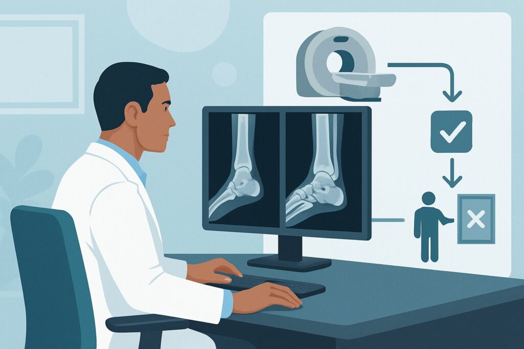
The results of ankle stress radiographs directly guide the next steps in patient management, creating a clear decision-making pathway. The goal is to triage patients who require surgical consultation from those who can be managed non-operatively.
If the stress views are positive, demonstrating objective evidence of instability (e.g., medial clear space widening greater than the superior clear space, or tibiofibular clear space >6 mm), the diagnosis of a significant syndesmotic and/or deltoid ligament injury is confirmed. This finding typically warrants an immediate orthopedic surgery consultation. The surgeon will then determine the need for operative stabilization, such as the placement of syndesmotic screws or a suture-button device to restore the integrity of the ankle mortise.
If the stress views are negative, meaning the joint remains stable under applied force, it suggests that while a ligamentous sprain is present, it is not severe enough to cause gross mechanical instability. However, if there remains a high clinical suspicion for a significant injury based on the examination (e.g., inability to bear weight, severe localized pain), the workup may proceed to the next appropriate study. In this case, an MRI of the ankle without IV contrast is the logical next step to directly visualize the ligaments, cartilage, and for occult bone injury.
If the study is indeterminate due to factors like patient guarding from pain preventing adequate stress, or if the measurements are borderline, the clinical pathway mirrors that of a negative study with high suspicion. Proceeding to MRI provides the definitive anatomic assessment needed to guide further treatment.
Pitfalls to Avoid (and When to Get Help)
Navigating the workup of suspected ankle instability requires attention to several common pitfalls to ensure an accurate and timely diagnosis.
- Inadequate Stress Application: A “negative” stress radiograph is only meaningful if sufficient stress was applied. If the patient’s pain and muscle guarding prevent an adequate examination, the result may be a false negative. Clear communication with the radiology department is key.
- Forgetting the Proximal Fibula: Always consider a Maisonneuve injury. Tenderness over the syndesmosis can be referred from a fracture of the proximal fibula. The initial radiographic series should include full-length tibia-fibula films if this injury is suspected.
- Misinterpreting Borderline Measurements: Radiographic measurements for instability have established cutoffs, but borderline cases exist. Correlating these findings with the physical exam is essential. An equivocal measurement in a patient with a grossly unstable-feeling ankle should be treated with a high degree of suspicion.
If there is any concern for a vascular injury, an open fracture, or compartment syndrome, this constitutes a surgical emergency. Escalate immediately to an orthopedic or vascular surgeon for emergent evaluation.
Related ACR Topics and Tools
This article focuses on a single, specific clinical scenario. For a comprehensive overview of all variants related to acute ankle trauma, or to explore the tools used to develop these recommendations, please refer to the following resources.
- For breadth across all scenarios in Acute Trauma to the Ankle, see our parent guide: Acute Trauma to the Ankle: ACR Appropriateness Decoded.
- To explore other clinical situations, use the ACR Appropriateness Criteria Lookup.
- For details on imaging techniques, consult the Imaging Protocol Library.
- To discuss radiation exposure with patients, the Radiation Dose Calculator can be a helpful aid.
Frequently Asked Questions
Why not just order an MRI for everyone with a suspected severe ankle sprain?
While MRI is excellent for anatomic detail and is rated ‘Usually Appropriate,’ stress radiographs are often preferred as the next step because they answer the immediate functional question: is the ankle mortise unstable? This is the key determination for surgical versus non-surgical management. Stress views are typically faster, more cost-effective, and more readily available in an acute setting. MRI is often reserved for cases where stress views are negative or equivocal but clinical suspicion remains high.
What specific measurements on a stress radiograph indicate syndesmotic instability?
On a mortise view with external rotation stress, several measurements are used. A tibiofibular clear space (the distance between the medial border of the fibula and the lateral border of the posterior tibial malleolus) greater than 6 mm is highly indicative of syndesmotic injury. Another sign is a tibiofibular overlap of less than 1 mm. A medial clear space wider than the superior clear space suggests associated deltoid ligament injury.
Can stress radiographs be performed on a child?
Yes, this guidance applies to children 5 years of age and older. The technique is similar, but it’s important to use pediatric-specific radiation dose protocols. The radiation dose from ankle stress views is very low (ACR RRL: ☢ <0.03 mSv for pediatrics). The primary challenge can be gaining the child's cooperation and ensuring pain is adequately managed to allow for a diagnostic study.
What if my facility does not perform stress radiographs?
If stress radiographs are not available, the ACR guidelines list both MRI without contrast and CT without contrast as ‘Usually Appropriate’ alternatives. In this situation, MRI is generally preferred as it provides superior evaluation of the ligaments and soft tissues. CT is more useful if an occult fracture is the primary concern. The choice depends on the specific clinical question and local resource availability.
Is there a role for weight-bearing radiographs in this scenario?
Weight-bearing radiographs can be considered a form of stress view and are very useful if the patient can tolerate them. If a patient can bear weight, obtaining weight-bearing AP and mortise views can sometimes reveal syndesmotic widening or talar tilt that was not apparent on non-weight-bearing films. However, many patients with acute, severe injuries cannot tolerate this, which is why manual or gravity stress views are often necessary.
Reviewed by Pouyan Golshani, MD, Interventional Radiologist — May 29, 2026