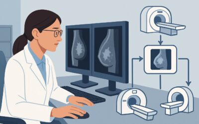
Should You Order Systemic Imaging for Distant Disease in Early-Stage Breast Cancer?

A 58-year-old woman presents to your clinic for treatment planning after a core biopsy confirmed a 1.8 cm, grade 2 invasive ductal carcinoma in her left breast. A physical exam is unremarkable, with no palpable axillary lymphadenopathy or skin changes. Her bloodwork, including liver function tests and alkaline phosphatase, is normal. You classify her with clinical stage I breast cancer and are now considering the next steps for staging. Is a full-body Positron Emission Tomography/Computed Tomography (PET/CT) scan or other systemic imaging necessary to evaluate for distant metastatic disease before proceeding with local therapy?
For this specific scenario—a newly diagnosed, clinical stage I-IIA breast cancer—the American College of Radiology (ACR) Appropriateness Criteria state that comprehensive imaging for distant disease, such as FDG-PET/CT, CT of the chest, abdomen, and pelvis, or a whole-body bone scan, is Usually Not Appropriate. This article details the evidence-based workflow for this common clinical decision point.
Who Fits This Clinical Scenario?
This guidance applies to patients with newly diagnosed invasive breast cancer (including invasive ductal carcinoma [IDC], invasive lobular carcinoma [ILC], or not otherwise specified [NOS]) who meet the criteria for early clinical stage I or IIA. These stages are generally defined by:
- Tumor size of 5 cm or less (T1 or T2)
- No or limited axillary lymph node involvement based on clinical exam (N0 or N1)
- No clinical or pathologic evidence of distant metastases (M0)
This workflow is specifically for the evaluation of distant disease. The evaluation for locoregional disease (the extent of cancer in the breast and nearby lymph nodes) is a separate and essential part of the workup, often involving dedicated breast and axillary imaging.
This article does not apply to patients who present with:
- More advanced disease (Stage IIB or III): Patients with larger tumors, extensive nodal involvement, or inflammatory breast cancer have a higher pre-test probability of distant metastases, and systemic staging is often warranted.
- Specific signs or symptoms of metastatic disease: A patient with new, persistent bone pain, abnormal liver function tests, or focal neurologic symptoms requires a targeted workup, regardless of their initial clinical stage.
- Evaluation for local recurrence: Surveillance imaging after initial treatment follows a different set of guidelines.
What Diagnoses Are You Working Up in This Scenario?
The primary goal of systemic staging is to identify or exclude distant metastases (M1 disease), which would fundamentally change the patient’s prognosis and treatment plan from curative-intent local therapy to systemic therapy for metastatic disease. In early-stage breast cancer, however, the pre-test probability of finding occult distant metastases is very low.
Exclusion of Distant Metastases: This is the principal objective. The key question is whether there is subclinical disease in the most common sites for breast cancer metastasis: bone, liver, lungs, and distant (non-axillary) lymph nodes. For patients with clinical stage I-IIA disease, the likelihood of identifying asymptomatic distant metastases is typically less than 5%. The clinical decision is whether the potential benefit of finding this rare event outweighs the harms of the imaging itself.
False-Positive Findings: A significant concern with performing high-sensitivity imaging in a low-prevalence population is the high rate of false-positive or incidental findings. A PET/CT or body CT might identify an indeterminate lung nodule, a benign liver hemangioma, or post-traumatic bony changes that are avid on a bone scan. These findings often trigger a cascade of further imaging, anxiety, and sometimes invasive biopsies for what ultimately proves to be a benign condition, delaying definitive cancer treatment.
Why Systemic Imaging for Distant Disease Is Usually Not Appropriate
For patients with newly diagnosed clinical stage I-IIA breast cancer, the ACR, along with other national guidelines (e.g., NCCN, ASCO), recommends against routine systemic staging for distant disease. The rationale is based on a careful balance of low diagnostic yield versus the potential harms of testing.
Studies such as FDG-PET/CT skull base to mid-thigh, CT chest abdomen pelvis with IV contrast, and Bone scan whole body are all rated as Usually Not Appropriate in this scenario. The reasons are multifaceted:
- Low Pre-Test Probability: As mentioned, the incidence of occult distant metastases in this patient population is very low. Performing sensitive tests in a low-prevalence setting dramatically increases the ratio of false-positive to true-positive results. The potential for patient harm from the workup of incidental findings outweighs the small chance of detecting true metastatic disease.
- Radiation Exposure: These staging studies involve significant ionizing radiation. A PET/CT or a contrast-enhanced CT of the chest, abdomen, and pelvis delivers a radiation dose of ☢☢☢☢ 10-30 mSv. A whole-body bone scan delivers ☢☢☢ 1-10 mSv. Avoiding this exposure is particularly important in a patient population with an excellent prognosis and long life expectancy following curative-intent therapy.
- Lack of Survival Benefit: Multiple studies have failed to demonstrate an overall survival benefit from performing routine systemic staging in asymptomatic, early-stage breast cancer patients. Treatment is not improved by finding asymptomatic metastases earlier in this specific context.
It is critical to distinguish this recommendation from the workup for locoregional disease. While systemic staging is not recommended, assessing the axillary lymph nodes is a standard and necessary part of the initial workup. An axillary ultrasound (US axilla) is often the first step in evaluating the nodal basin, though its appropriateness is formally addressed under the ACR scenario for locoregional, not distant, disease evaluation.
What’s Next? Downstream Workflow After Deciding Against Systemic Imaging
After a thorough history and physical exam confirm the patient is asymptomatic and fits the stage I-IIA criteria, the decision is made to forgo systemic staging. The clinical workflow then proceeds directly to planning for definitive local and systemic therapy.
- If the decision is NO systemic imaging: The patient’s care proceeds to the multidisciplinary tumor board for discussion. The plan will focus on locoregional management (e.g., lumpectomy or mastectomy, sentinel lymph node biopsy, and possibly radiation) and adjuvant systemic therapy (e.g., chemotherapy, endocrine therapy, targeted therapy) based on the tumor’s pathology and biomarker status (ER, PR, HER2).
- If the patient develops symptoms: If at any point before or during treatment the patient develops new, concerning symptoms (e.g., localized bone pain, unexplained cough, jaundice), the “no imaging” decision should be immediately reconsidered. At that point, a targeted imaging study (e.g., MRI of the spine for back pain, CT chest for cough) becomes appropriate to evaluate for a specific metastatic site.
- If pathology is upstaged: If surgical pathology (e.g., from sentinel node biopsy) reveals a higher disease burden than expected, upstaging the patient to stage IIB or III, then post-operative systemic staging may become appropriate. This represents a change in the clinical scenario.
Pitfalls to Avoid (and When to Get Help)
Navigating the staging of early breast cancer requires avoiding several common pitfalls. Be mindful of these to ensure evidence-based, patient-centered care.
- The “One-Size-Fits-All” Workup: Avoid reflexively ordering a PET/CT or other systemic imaging for every new breast cancer diagnosis. Staging should be tailored to the clinical stage and individual patient risk.
- Ignoring Clinical Red Flags: The recommendation against routine imaging applies only to asymptomatic patients. Do not dismiss new, persistent, or localized symptoms that could indicate metastatic disease.
- Confusing Locoregional vs. Distant Staging: The decision to omit systemic staging for distant disease does not mean omitting locoregional staging. A thorough clinical exam and appropriate axillary imaging (typically ultrasound) remain critical.
- Over-reliance on Tumor Markers: Routine measurement of serum tumor markers like CA 15-3 or CA 27.29 is not recommended for staging. These markers lack the sensitivity and specificity for this purpose and should not be used to trigger staging imaging in an otherwise asymptomatic, early-stage patient.
If you are uncertain whether a patient’s symptoms or high-risk tumor biology (e.g., triple-negative, high grade) warrant an exception, discussion with a multidisciplinary team, including medical oncology and breast radiology, is the best next step.
Related ACR Topics and Tools
This article covers a single, focused clinical scenario. For a broader view of the topic and for tools to help with adjacent clinical questions, the following resources are available.
- For breadth across all scenarios in Imaging of Invasive Breast Cancer, see our parent guide: Imaging of Invasive Breast Cancer: ACR Appropriateness Decoded.
- To look up other clinical variants or ACR recommendations, use the ACR Appropriateness Criteria Lookup.
- For detailed procedural techniques on recommended studies, consult the Imaging Protocol Library.
- To discuss radiation exposure with patients, the Radiation Dose Calculator can help quantify and contextualize the dose from various exams.
Frequently Asked Questions
Does the recommendation change if the histology is Invasive Lobular Carcinoma (ILC)?
For early-stage (I-IIA) disease, the recommendation generally does not change based on histology alone. While ILC can have a different pattern of metastasis and may be less FDG-avid on PET scans, the overall risk of distant metastases in asymptomatic, early-stage ILC is still too low to justify routine systemic staging. The same principles of low diagnostic yield and high risk of false positives apply.
Are there any exceptions for Stage IIA patients where systemic imaging might be considered?
While guidelines recommend against routine staging for all Stage I-IIA patients, some clinicians may consider it for the highest-risk subset, such as those with T2N1 disease and aggressive biology (e.g., triple-negative or high-grade HER2-positive). However, this is not a universal recommendation, and the decision should be made on a case-by-case basis, ideally in a multidisciplinary discussion, acknowledging the limited evidence for a survival benefit.
What should I do if my patient is very anxious and requests a ‘full body scan’ for peace of mind?
This is a common and understandable request. The best approach is shared decision-making. Acknowledge the patient’s anxiety and clearly explain the rationale against routine scanning: the very low chance of finding anything, the high chance of false positives leading to more tests and delays, and the unnecessary radiation exposure. Framing the decision as an evidence-based way to avoid harm and proceed more quickly to effective treatment can be helpful.
You said not to perform systemic imaging. Does this include axillary staging?
No, this is a critical distinction. The recommendation against systemic imaging applies to the search for *distant* metastases (in bones, liver, lungs, etc.). *Locoregional* staging, which includes a clinical examination of the axilla and typically an axillary ultrasound to evaluate the lymph nodes, is an essential part of the initial workup for nearly all patients with invasive breast cancer.
What if the alkaline phosphatase is mildly elevated without any other symptoms?
An isolated, mildly elevated alkaline phosphatase can be a challenging scenario. It can be a sign of bone or liver metastases, but it also has many benign causes. If the elevation is minimal and the patient is completely asymptomatic with an otherwise normal physical exam and liver function tests, most guidelines would still recommend against a full systemic imaging workup. The decision may be individualized, possibly starting with a fractionated alkaline phosphatase to determine its origin (bone vs. liver) before proceeding to imaging.
Reviewed by Pouyan Golshani, MD, Interventional Radiologist — May 30, 2026