
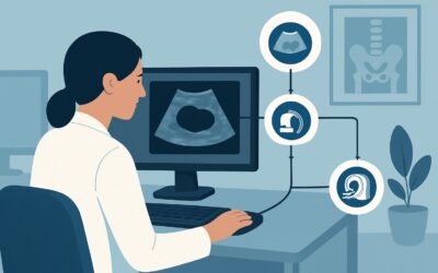
What Is the Best Initial Imaging for a Suspected Adnexal Mass in Pregnancy?
ACR-guided imaging workflow for Female. Clinically suspected adnexal mass, no acute symptoms. Pregnant. Initial imaging. (Clinically Suspected Adnexal Mass
ACR-guided imaging workflow for Female. Clinically suspected adnexal mass, no acute symptoms. Pregnant. Initial imaging. (Clinically Suspected Adnexal Mass

ACR-guided imaging workflow for Adult female. Adnexal mass, suspicious for malignancy on pelvic US, no acute symptoms. Premenopausal or postmenopausal. Nex

ACR-guided imaging workflow for Adult female. Adnexal mass, indeterminate on initial pelvic US, no acute symptoms. Postmenopausal. Follow-up imaging. (Clin
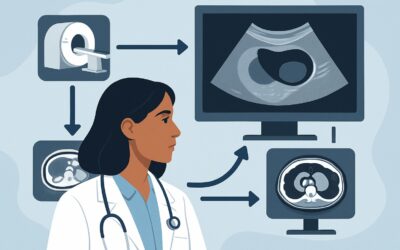
ACR-guided imaging workflow for Adult female. Adnexal mass, indeterminate on initial pelvic US, no acute symptoms. Premenopausal. Follow-up imaging. (Clini

ACR-guided imaging workflow for Adult female. Adnexal mass, indeterminate on initial pelvic US, no acute symptoms. Premenopausal or postmenopausal. Next im
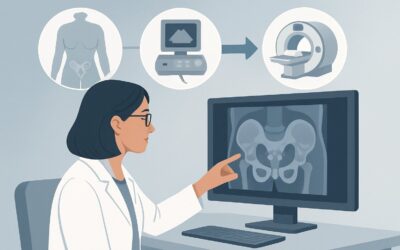
ACR-guided imaging workflow for Adult female. Clinically suspected adnexal mass, no acute symptoms. Premenopausal or postmenopausal. Initial imaging. (Clin
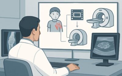
ACR-guided imaging workflow for Child. Established diagnosis of vascular malformation presenting with new or persistent signs or symptoms. Initial imaging.
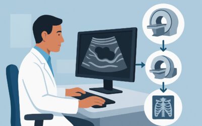
ACR-guided imaging workflow for Child. Ultrasound features raise suspicion for vascular malformation. Next imaging study. (Soft Tissue Vascular Anomalies:
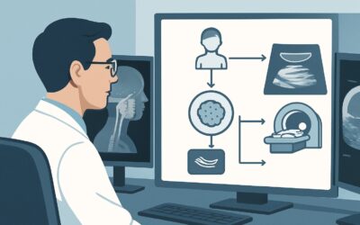
ACR-guided imaging workflow for Child. Clinical signs or symptoms of vascular anomaly (tumor or malformation) not suggesting infantile hemangioma. Initial
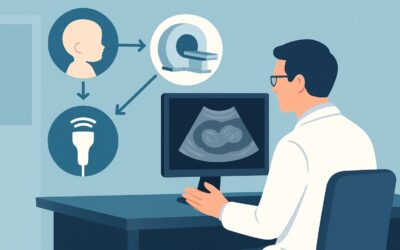
ACR-guided imaging workflow for Infant. Multiple cutaneous infantile hemangiomas, screening for infantile hepatic hemangiomas. Initial imaging. (Soft Tissu