

Hip X-Ray — Dictation, Appropriateness, and Dose for Residents
Plain radiograph of hip — first-line for fracture, dislocation, arthritis. AP pelvis + frog-leg lateral or cross-table lateral standard.
Plain radiograph of hip — first-line for fracture, dislocation, arthritis. AP pelvis + frog-leg lateral or cross-table lateral standard.

Standard 3-view radiographic series of the hand or wrist (PA, lateral, oblique) for fracture, dislocation, arthritis, foreign body, and post-operative…
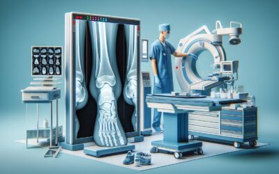
Plain radiograph of ankle and foot — first-line per Ottawa Ankle and Foot Rules for trauma.

Plain radiograph of the shoulder — Grashey AP, axillary, outlet (scapular Y) views. First-line for fracture, dislocation, AC joint, and acromial…

Multi-view radiographic series of the knee (AP, lateral, sunrise/skyline ± Rosenberg or notch) for evaluation of fracture, dislocation, osteoarthritis…

Multi-view radiographic series of the cervical spine (AP, lateral, open-mouth odontoid; ± obliques and flexion-extension) for evaluation of trauma,…

AP and lateral radiographic views of the lumbar spine for evaluation of low back pain, vertebral fracture, alignment, scoliosis, and post-operative spine….

Plain radiograph of the abdomen — supine and/or upright views. Limited modern role; mainly for tube/line position, foreign body, gross obstruction, and…

Single-view AP chest radiograph performed at the bedside (ICU, ED, OR) for evaluation of acute cardiopulmonary processes, line/tube placement, and serial…

Two-view (PA + lateral) plain radiograph of the chest — the most commonly performed imaging study globally and the workhorse for pulmonary, cardiac,…