US MSK Shoulder — Dictation, Appropriateness, and Dose for Residents
1. The High-Yield Shoulder Ultrasound: A Systematic Approach
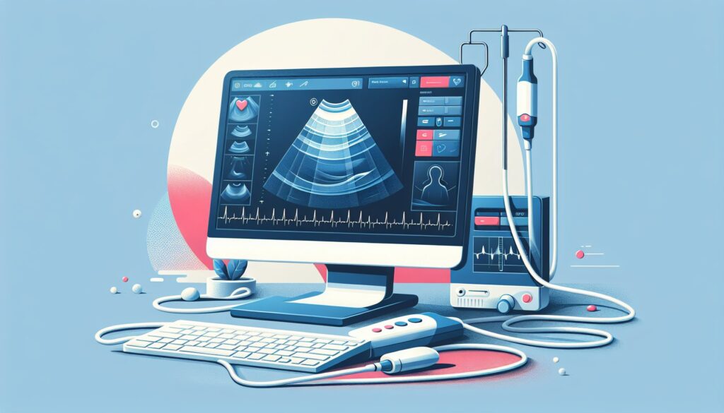
You’re on your MSK rotation, and the list is full of outpatient shoulder ultrasounds. The referring orthopedic surgeon wants to know if the patient’s positive drop arm test is from a full-thickness tear before committing to a scope. This isn’t a passive CT where the images just show up; it’s an operator-dependent exam where you drive the transducer. Miss a position, and you might miss the tear. Your attending expects a systematic evaluation of every single tendon, the AC joint, and a dynamic assessment for impingement. It’s a lot to keep straight when you’re trying to be efficient.
When I was a resident, I taped a diagram of the 6 standard shoulder positions to the ultrasound machine. There’s no shame in a good checklist. The key is turning that systematic scan into a clean, structured report that answers the clinical question without ambiguity. For more high-yield guides like this, check out the free trainee calculators and references we’ve put together.
2. What a Musculoskeletal Shoulder Ultrasound Covers and What Attendings Look For
A shoulder ultrasound is the workhorse for evaluating rotator cuff and biceps pathology. Its major advantage over MRI is the ability to perform dynamic imaging—watching the tendons and bursae move in real-time as the patient repositions their arm. This is crucial for assessing issues like subacromial impingement.
Your attending expects a comprehensive report that methodically addresses each component of the shoulder cuff and adjacent structures. The goal is to answer these key clinical questions:
- Is there a full-thickness or partial-thickness rotator cuff tear, particularly of the supraspinatus?
- Are there signs of calcific tendinopathy, and if so, what type?
- Is the long head of the biceps tendon normal in appearance and located correctly within the bicipital groove?
- Is there evidence of subacromial-subdeltoid bursitis?
- What is the status of the acromioclavicular (AC) joint?
- Can dynamic impingement be demonstrated?
This study is the first choice for shoulder pain after trauma, suspected cuff tears, localizing calcific deposits for intervention, and guiding injections. It’s less effective for deep structures like the labrum or for evaluating bone pathology, where MRI or CT are superior.
3. Radiology Report Template for Musculoskeletal Shoulder Ultrasound (Rotator Cuff)
This template provides a reliable framework. The key is to be systematic in your findings, addressing each structure you evaluated, even if it’s normal. This tells the referring clinician you looked for specific pathology and didn’t find it.
Technique
Real-time grayscale and color Doppler ultrasound evaluation of the right/left shoulder was performed using a high-frequency linear transducer. A standardized protocol was used to evaluate the rotator cuff tendons, long head of the biceps tendon, acromioclavicular joint, and subacromial-subdeltoid bursa. Dynamic imaging was performed.
Findings
Long Head of Biceps Tendon: The long head of the biceps tendon is normal in caliber and echotexture, and is located within the bicipital groove. No significant tenosynovial fluid. [OR: Describe tendinosis, partial/full tear, dislocation, or tenosynovial fluid.]
Subscapularis Tendon: The subscapularis tendon is intact at its insertion on the lesser tuberosity, with a normal fibrillar pattern. [OR: Describe tendinosis, calcifications, or partial/full-thickness tear.]
Supraspinatus Tendon: The supraspinatus tendon is intact at its insertion on the greater tuberosity. Normal echotexture. [OR: Describe tendinosis, calcifications, or partial/full-thickness tear specifying location (articular surface, bursal surface, intrasubstance) and size in two dimensions.]
Infraspinatus and Teres Minor Tendons: The infraspinatus and teres minor tendons are intact. Normal echotexture. [OR: Describe tendinosis, calcifications, or tears.]
Subacromial-Subdeltoid Bursa: No significant bursal fluid or synovial thickening. [OR: Distended with fluid, consistent with bursitis.]
Acromioclavicular (AC) Joint: Unremarkable. No significant capsular thickening or joint effusion. [OR: Degenerative changes with osteophytes and capsular hypertrophy.]
Posterior Glenohumeral Joint: No joint effusion. The posterior labrum is grossly intact. [Evaluation of the labrum is limited on ultrasound.]
Dynamic Assessment: During dynamic abduction, there is no evidence of supraspinatus tendon “humping” or impingement beneath the acromion. [OR: There is bunching of the supraspinatus tendon and subacromial bursa beneath the acromion, consistent with subacromial impingement.]
Impression
- [Normal study OR specific findings, e.g., “Full-thickness tear of the anterior supraspinatus tendon measuring X x Y cm.”]
- [Secondary findings, e.g., “Mild subacromial-subdeltoid bursitis.”]
- [Other relevant findings, e.g., “Degenerative changes of the acromioclavicular joint.”]
4. Where to Find More Free Radiology Report Templates
Building your own library of high-quality templates is one of the best things you can do during training. While you’ll develop your own style, starting with a solid, community-vetted foundation is key. Beyond your institution’s shared macros, two great free repositories exist that are worth bookmarking.
- RadReport.org: Curated by the Radiological Society of North America (RSNA), this is a comprehensive library of peer-reviewed templates covering nearly every modality and subspecialty. They are designed for structured reporting and are a go-to resource.
- Radiology Templates (AU): This is an excellent, user-friendly site maintained by Australian radiologists. It offers clean, practical templates that are easy to adapt for your own use.
5. From Free-Form Dictation to a Flawless Structured Report
The real challenge on a busy service isn’t just finding the pathology; it’s communicating it clearly and quickly. Standard templates are a great start, but they can feel rigid when you have multiple positive findings. You end up jumping around, deleting negative statements, and manually filling in measurements. This is where AI-assisted reporting can streamline your workflow.
Instead of clicking through a rigid template, you can simply dictate your positive findings in free form—”full-thickness tear of the supraspinatus at the footprint measuring 1.2 by 0.8 cm with associated subacromial bursitis.” The software then parses this and generates a perfectly structured report. Tools like GigHz Precision AI are designed for this, using pre-loaded templates from the American College of Radiology (ACR) and Society of Interventional Radiology (SIR). It also helps surface the appropriate Clinical Decision Support (CDS) frameworks, like LI-RADS or Bosniak, when they’re needed, ensuring your reports meet attending and payer expectations every time.
6. When Should You Order a Shoulder Ultrasound? ACR Appropriateness Criteria
Understanding when a shoulder ultrasound is the right first test is crucial. The American College of Radiology (ACR) provides evidence-based guidelines to help with this decision. For the common indication of non-traumatic shoulder pain, the guidance is clear.
According to the ACR Appropriateness Criteria for Shoulder Pain-Atraumatic, a shoulder ultrasound “may be appropriate” as an initial imaging study. It is particularly useful for evaluating the rotator cuff tendons and for dynamic assessment of impingement. However, for a more comprehensive evaluation of the shoulder, including the labrum, cartilage, and bone marrow, an MRI of the shoulder without contrast is “usually appropriate.” If there’s a specific concern for labral pathology or instability, an MR arthrogram is the preferred study.
Essentially, ultrasound is an excellent, accessible, and cost-effective first-line tool for cuff and biceps pathology. If the clinical picture is more complex or if the ultrasound is inconclusive, MRI is the logical next step.
7. How Much Radiation Does a Shoulder Ultrasound Deliver?
One of the primary advantages of musculoskeletal ultrasound is its safety profile. A shoulder ultrasound delivers an effective radiation dose of 0 mSv. It uses sound waves, not ionizing radiation, to create images, posing no radiation risk to the patient.
This makes it an ideal imaging modality for young patients, pregnant patients, and for situations requiring repeated follow-up imaging or image guidance for procedures. The absence of radiation is a significant benefit compared to other modalities like CT or even standard radiographs.
| Imaging Study | Effective Radiation Dose | Comparison |
|---|---|---|
| Shoulder Ultrasound | 0 mSv | No radiation |
| Shoulder X-ray (2 views) | ~0.01 mSv | Less than 1 day of natural background radiation |
| Shoulder CT | ~2-4 mSv | Equivalent to ~1 year of natural background radiation |
8. Musculoskeletal Shoulder Ultrasound Protocol — Positions, Transducer, and Dynamic Maneuvers
A successful shoulder ultrasound relies on a standardized, multi-position protocol to ensure all relevant structures are visualized. The patient is typically seated on a rotating stool to facilitate easy repositioning. A high-frequency (10-15 MHz) linear transducer is used to provide high-resolution images of these superficial structures.
The following table outlines the standard 6-position protocol that serves as the foundation for a comprehensive exam. Following this sequence ensures nothing is missed, from the biceps tendon anteriorly to the posterior cuff.
| Position | Patient Maneuver | Key Structures Visualized |
|---|---|---|
| 1. Biceps Tendon | Palm up on thigh | Long head of biceps tendon in bicipital groove, transverse subscapularis |
| 2. Subscapularis | External rotation | Subscapularis tendon insertion on lesser tuberosity |
| 3. Supraspinatus | Modified Crass position (hand on opposite buttock) | Supraspinatus tendon footprint (longitudinal and transverse views) |
| 4. Infraspinatus/Teres Minor | Hand on opposite shoulder | Posterior cuff tendons inserting on greater tuberosity |
| 5. AC Joint | Neutral position | Acromioclavicular joint, distal clavicle, acromion |
| 6. Posterior Joint | Hand on opposite shoulder | Posterior glenohumeral joint for effusion, posterior labrum |
Common protocol pitfalls: The most critical step is achieving an adequate modified Crass position. If the patient can’t get their hand far enough behind their back, the supraspinatus tendon will remain tucked under the acromion, leading to a non-diagnostic study. In these cases, document the limitation and consider alternative views or recommending MRI.
9. The 3-Months-Free Offer for Residents and Fellows
3+ months free for radiology residents and fellows
We want to help you look like a rockstar on your reports. GigHz Precision AI is designed to help you dictate positive findings in free form, then automatically generate a clean, structured draft using ACR and SIR templates. The appropriate clinical decision support fires automatically, so you’re not hunting for calculators mid-read. All we ask in return is your feedback so we can keep improving the product for trainees.
Signup is simple. There’s no credit card required and no long forms. To get set up, just provide these three items:
- Your PGY year (e.g., PGY-2, PGY-4)
- Your training type (radiology residency or fellowship specialty)
- Your training program / hospital name
Ready to give it a try? Apply for the residents free-access program and we’ll get you set up.
10. Frequently Asked Questions
Is GigHz Precision AI HIPAA-compliant?
Yes. The platform is designed for de-identified workflows by default. No Protected Health Information (PHI) is required to use the tool for generating structured report text from your findings. It operates on the text of your dictation, not on patient-linked data from the EMR or PACS.
Do I need my hospital’s IT department to set this up?
No. GigHz Precision AI is browser-based and requires no local software installation or special permissions. It works on any modern web browser, including the one on your call-room workstation or personal iPad.
How does this work with PowerScribe or other dictation systems?
It works alongside your existing dictation system. You can dictate your findings, use the tool to generate the structured report text, and then copy/paste that clean text into your PACS/RIS for final signature. It’s a workflow enhancement, not a replacement for your core dictation software.
Can I use this on my phone or iPad?
Yes, the platform is fully responsive and works well on tablets like the iPad, which is common in reading rooms. While it functions on a mobile phone, the screen size is better suited for a tablet or desktop for reviewing and editing reports.
Can I customize the templates?
The core templates are based on official ACR and SIR guidelines to ensure compliance and standardization. However, the output is fully editable text, so you can easily tweak the final report to match your preferred phrasing or your attending’s specific preferences before signing.
What happens after my residency or fellowship ends?
The free access program is specifically for trainees. After you graduate, you can transition to a standard plan for practicing radiologists. We have straightforward, transparent pricing for individual physicians and groups.
Free GigHz Tools That Pair With This Article
Three free tools that complement the material above:
- Imaging Appropriateness Selector — Type an indication or clinical scenario in plain language and get the imaging studies the ACR rates for it, with adult and pediatric radiation levels. Built directly from 297 ACR topics, 1,336 clinical variants, and 15,823 procedure ratings.
- GigHz Imaging Protocol Library — A searchable library of 131 imaging protocols with the physics specs surfaced and the matching ACR Appropriateness Criteria alongside. Plain-English narratives readable in 60 seconds, organized by modality.
- GigHz Radiation Dose Calculator — Pick the imaging studies a patient has had and see total dose in millisieverts (mSv) with comparisons to natural background radiation, transatlantic flights, and chest X-rays. Useful for shared decision-making.
Last reviewed by Pouyan Golshani, MD — 2026-08-04.
Reviewed by Pouyan Golshani, MD, Interventional Radiologist — August 4, 2026