What Imaging Best Assesses Esophageal Cancer Response During Treatment?

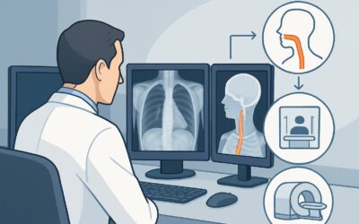
A 64-year-old male with locally advanced adenocarcinoma of the distal esophagus is midway through his neoadjuvant chemoradiation protocol. His oncologist needs to assess the tumor’s response to decide whether to continue the current regimen or pivot strategy before his planned esophagectomy. The clinical question is pointed: is the therapy working, and how can imaging provide a definitive answer before anatomical changes are obvious? This scenario requires a modality that can evaluate metabolic activity, not just tumor size. For assessing esophageal cancer during treatment, the American College of Radiology (ACR) Appropriateness Criteria rate FDG-PET/CT skull base to mid-thigh as Usually Appropriate.
Who Fits This Clinical Scenario?
This guidance applies specifically to patients with a confirmed diagnosis of esophageal cancer who are currently undergoing neoadjuvant therapy (chemotherapy, radiation, or both) prior to planned definitive treatment, which is often surgery. The primary goal of imaging in this context is to assess interim treatment response, which can be a powerful prognostic indicator and may guide subsequent management decisions.
This workflow is distinct from other common clinical timepoints in esophageal cancer care. It does not apply to:
- Initial Pretreatment Staging: Patients who have a new diagnosis but have not yet started therapy require a different imaging workup to establish the baseline extent of disease. This is covered in the ACR variant for initial staging.
- Post-Treatment Surveillance: Patients who have completed all planned therapy and are being monitored for recurrence without specific symptoms or signs fall under routine follow-up guidelines.
- Suspected Recurrence: Patients who have completed therapy and now present with new symptoms (like dysphagia, weight loss, or pain) concerning for recurrent disease require a workup focused on identifying and characterizing the new abnormality.
The key differentiator for this scenario is the timing: the patient is actively on treatment, and the imaging is intended to inform the immediate next steps of that ongoing therapeutic course.
What Diagnoses Are You Working Up in This Scenario?
In the mid-treatment setting, imaging is not for initial diagnosis but for evaluating the biologic effects of therapy. The “differential” is a spectrum of treatment responses and related complications.
Favorable Metabolic Response
This is the primary goal. A significant reduction in the tumor’s metabolic activity, measured by Fluorodeoxyglucose (FDG) uptake on a PET scan, indicates that the cancer cells are responding to chemotherapy and/or radiation. This finding is a strong positive prognostic indicator and supports continuing the current treatment plan toward definitive surgery.
Progressive Metabolic Disease
The most concerning finding is stable or, worse, increasing metabolic activity in the primary tumor or the appearance of new metastatic sites. This suggests the cancer is resistant to the current neoadjuvant regimen. Identifying this early allows the multidisciplinary team to consider altering the treatment strategy, potentially avoiding the morbidity of an ineffective therapy and a futile major surgery.
Stable Disease
The tumor may show little to no change in metabolic activity or size. This indeterminate result presents a clinical challenge, suggesting a suboptimal response and prompting a discussion about the risks and benefits of proceeding with the original plan versus modifying treatment.
Treatment-Related Inflammation (Esophagitis)
Radiation therapy invariably causes inflammation of the surrounding healthy esophageal tissue, a condition known as radiation esophagitis. This inflammation is also metabolically active and will demonstrate FDG uptake on a PET scan. Differentiating this benign inflammatory uptake from residual malignant tumor is a critical interpretive challenge and a major reason why expertise in reading these scans is essential.
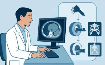
Why Is FDG-PET/CT the Recommended Study for Assessing Treatment Response?
The ACR designates FDG-PET/CT skull base to mid-thigh as Usually Appropriate because it uniquely combines functional (metabolic) data with anatomic localization, providing the most sensitive assessment of early treatment response.
A tumor’s metabolic response often precedes any measurable change in its size. A conventional CT scan might show a tumor that is unchanged in dimension, while a PET scan reveals that its metabolic engine has been shut down by therapy. This early insight is invaluable. Studies have shown that the degree of reduction in FDG uptake on an interim PET/CT is a powerful predictor of pathologic response and overall survival.
Why are other common modalities rated lower for this specific task?
- CT chest abdomen pelvis with IV contrast is rated Usually Not Appropriate. While CT provides excellent anatomic detail, it cannot reliably distinguish viable tumor from post-treatment scarring, edema, or inflammation. A non-enlarging but metabolically active tumor would be missed, and a shrinking but still viable tumor could be misinterpreted.
- Fluoroscopy upper GI series is also Usually Not Appropriate for this indication. A barium esophagram is excellent for evaluating luminal patency and swallowing function but provides no information about tumor metabolism or nodal/distant metastatic disease.
The recommended study, FDG-PET/CT, involves a significant radiation dose (ACR Relative Radiation Level ☢☢☢☢), but this is justified by the high-stakes clinical question being answered. The information gained can prevent a patient from undergoing a major esophagectomy if the tumor is not responding or has already metastasized, a benefit that far outweighs the radiation risk in this patient population.
What’s Next After FDG-PET/CT? Downstream Workflow
The results of the interim FDG-PET/CT should be discussed in a multidisciplinary tumor board to guide the next steps in management. The downstream workflow depends directly on the findings.
If the study shows a good metabolic response:
This is a positive prognostic sign. The patient typically completes the planned course of neoadjuvant chemoradiation. The next step is usually pre-surgical planning, followed by esophagectomy as originally intended. The imaging provides confidence that the patient is on the right track.
If the study shows progressive disease (metabolic or anatomic):
This is a critical alert. The finding of treatment resistance or new metastases necessitates an immediate re-evaluation of the treatment plan. The team may consider switching to a different systemic therapy regimen, adding or modifying radiation, or, in the case of new distant metastases, shifting the goal of care from curative to palliative. Proceeding with a major surgery in the face of progressive disease is often futile and exposes the patient to significant risk without benefit.
If the study is indeterminate or shows stable disease:
This is the most complex scenario. The findings may be ambiguous due to significant treatment-related esophagitis confounding the interpretation. The next step often involves correlation with other clinical data. An esophagogastroduodenoscopy (EGD) with biopsy may be performed to pathologically assess the residual tissue. The tumor board may decide to continue with the planned therapy and surgery, acknowledging the uncertain prognosis, or consider a modification to the treatment plan.
Pitfalls to Avoid (and When to Get Help)
Several common pitfalls can compromise the utility of mid-treatment imaging in esophageal cancer. Awareness of these issues is key to obtaining an accurate and actionable result.
- Improper Timing: Performing the PET/CT too soon after a cycle of chemotherapy or, more importantly, too soon after the completion of radiation therapy can lead to false-positive results from intense inflammatory uptake. A sufficient washout period (typically at least 4-6 weeks post-radiation) is crucial if the scan is for post-treatment assessment, though mid-treatment protocols vary.
- Ignoring Blood Glucose Levels: Hyperglycemia competitively inhibits FDG uptake by tumor cells, potentially causing a false-negative scan. Always ensure the patient is appropriately fasted and that their blood glucose is within an acceptable range (e.g., <150-200 mg/dL) before the radiotracer is injected.
- Overlooking Esophagitis: Failing to consider radiation-induced esophagitis as a cause for FDG uptake can lead to misinterpretation of residual disease. The pattern of uptake (diffuse and linear vs. focal and nodular) and correlation with the CT portion of the scan are key to differentiation.
If the imaging results are equivocal or conflict with the clinical picture, immediate escalation to the multidisciplinary tumor board is the most appropriate next step. Integrating insights from medical oncology, radiation oncology, surgery, and radiology is essential for optimal decision-making.
Related ACR Topics and Tools
For a comprehensive overview of all clinical scenarios related to imaging esophageal cancer, from initial diagnosis to long-term follow-up, please consult our parent topic guide. It provides a breadth of information that complements this in-depth article.
- For breadth across all scenarios in Staging and Follow-up of Esophageal Cancer, see our parent guide: Staging and Follow-up of Esophageal Cancer: ACR Appropriateness Decoded.
- To explore other clinical situations, use the ACR Appropriateness Criteria Lookup.
- For details on imaging techniques, visit the Imaging Protocol Library.
- To discuss radiation exposure with patients, the Radiation Dose Calculator can help frame the conversation.
Frequently Asked Questions
Why not just use a diagnostic CT with contrast instead of a PET/CT?
A standard CT scan primarily shows anatomy and tumor size. During treatment, a tumor may not shrink significantly but can still be responding metabolically. Conversely, inflammation and scarring can mimic residual tumor on CT. FDG-PET/CT is superior because it assesses the tumor’s metabolic activity, providing a much earlier and more accurate indicator of treatment response than size criteria alone.
How long after radiation therapy should we wait to perform the PET/CT?
The timing is critical. Radiation causes intense inflammation (esophagitis), which is also FDG-avid and can be mistaken for cancer. For a post-treatment response assessment, it is generally recommended to wait at least 4-6 weeks, and sometimes up to 12 weeks, after the completion of radiation therapy to allow this inflammation to subside. For an interim scan during a course of chemoradiation, the protocol should be determined by the institutional tumor board, but the timing relative to the last radiation dose is a key factor in interpretation.
What if the patient’s blood sugar is high on the day of the PET/CT scan?
High blood glucose levels (hyperglycemia) will lead to a poor-quality, often non-diagnostic, PET scan. Glucose in the blood competes with the FDG radiotracer for uptake into cells, including cancer cells. This can make a tumor appear falsely negative or have artificially low activity. Most imaging centers have a strict glucose cutoff (e.g., 150-200 mg/dL). If the patient’s level is too high, the scan should be rescheduled.
Can FDG-PET/MRI be used instead of FDG-PET/CT in this scenario?
FDG-PET/MRI is rated as *May be appropriate* by the ACR for this indication. It offers the same metabolic data from PET with the superior soft-tissue contrast of MRI, which can be advantageous for evaluating local tumor invasion. However, it is less widely available, more expensive, and takes longer to perform than PET/CT. For the primary questions of metabolic response and detecting distant metastases, PET/CT remains the established standard of care.
What is the next step if the PET/CT shows a complete metabolic response?
A complete metabolic response is an excellent prognostic sign. The patient should continue with the planned course of therapy, which typically involves proceeding to surgery (esophagectomy) after the neoadjuvant phase is complete. This finding gives the surgical team confidence that the preoperative therapy was effective and that the patient is optimized for a curative-intent operation.
Reviewed by Pouyan Golshani, MD, Interventional Radiologist — May 30, 2026