What Imaging Is Best for a Child with a Suspected Secondary Headache?

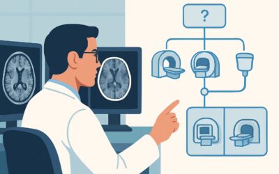
An 8-year-old boy presents to your clinic with a six-week history of progressively worsening morning headaches, occasionally associated with nausea. Over the past week, his parents have noticed some mild clumsiness and imbalance. On examination, you note subtle dysmetria on finger-to-nose testing. This constellation of “red flag” symptoms raises concern for a secondary cause of his headaches, distinct from a typical primary migraine or tension-type headache. The immediate clinical question is which imaging study to order to investigate for an underlying structural cause. This article provides a focused workflow for this specific scenario, explaining why the American College of Radiology (ACR) rates MRI head without and with IV contrast as Usually appropriate.
Who Fits This Clinical Scenario?
This guidance applies to a pediatric patient (child or adolescent) presenting with a headache that has features suspicious for a secondary, or underlying structural, cause. These “red flag” features are critical to recognize and differentiate this scenario from the more common primary headache syndromes. Key inclusion criteria for this workflow include:
- New or worsening headache pattern: A headache that is new, progressively increasing in frequency or severity, or has changed character significantly.
- Associated neurological signs or symptoms: Any focal neurologic deficit, such as weakness, sensory changes, ataxia, cranial nerve palsies, or changes in coordination.
- Signs of increased intracranial pressure: Headaches that are worse in the morning, wake the child from sleep, are exacerbated by coughing or Valsalva maneuvers, or are associated with vomiting. Papilledema on funduscopic exam is a classic, albeit late, sign.
- Systemic symptoms: Unexplained fever, weight loss, or other constitutional symptoms in conjunction with the headache.
This workflow is distinct from other pediatric headache scenarios. It does not apply to a child with a classic primary headache (e.g., migraine with aura, tension-type headache) without red flag features. It also differs from the workup for a sudden, severe “thunderclap” headache, which prioritizes ruling out subarachnoid hemorrhage, or a headache directly attributed to a known recent infection or significant head trauma, each of which follows a separate diagnostic pathway.
What Diagnoses Are You Working Up in This Scenario?
When ordering imaging for a suspected secondary headache in a child, the goal is to identify or exclude serious intracranial pathology. The differential diagnosis is broad, but the most consequential possibilities drive the choice of imaging modality. These include intracranial masses, hydrocephalus, and vascular abnormalities.
Intracranial Mass: This is often the primary concern. In children, approximately two-thirds of brain tumors are located in the posterior fossa (cerebellum and brainstem). Common pediatric tumors like medulloblastoma, pilocytic astrocytoma, and ependymoma typically arise here and present with symptoms of increased intracranial pressure and cerebellar dysfunction (e.g., ataxia, dysmetria), mirroring the clinical vignette. These tumors often enhance with contrast, a key feature for their detection and characterization.
Hydrocephalus: This refers to the abnormal accumulation of cerebrospinal fluid (CSF) within the ventricles. It can be obstructive, caused by a mass or stenosis blocking CSF flow (e.g., aqueductal stenosis), or communicating, resulting from impaired CSF absorption. Hydrocephalus is a direct cause of increased intracranial pressure and headache. Imaging is essential to identify the presence and level of obstruction.
Vascular Malformations: Less common but critical to identify, conditions like arteriovenous malformations (AVMs), cavernous malformations, or dural arteriovenous fistulas can present with headache. While their classic presentation is often hemorrhage, headache can be a sentinel symptom. These lesions have characteristic appearances on specific MRI sequences.
Inflammatory or Demyelinating Conditions: Though less frequent, conditions like acute disseminated encephalomyelitis (ADEM) or other central nervous system inflammatory processes can present with headache and multifocal neurologic deficits. These pathologies are characterized by areas of inflammation that are best visualized with contrast-enhanced MRI.
Why Is MRI Head Without and With IV Contrast the Recommended Study?
The ACR designates MRI head without and with IV contrast as Usually appropriate for this clinical scenario because it provides the highest diagnostic yield for the key differential diagnoses while avoiding ionizing radiation. The superior soft-tissue contrast of Magnetic Resonance Imaging (MRI) is unmatched for evaluating the brain parenchyma, especially the posterior fossa, where pediatric tumors are common and CT imaging is often limited by beam-hardening artifact from the surrounding bone.
The rationale for this specific protocol involves several key components:
- Sensitivity for Pathology: Non-contrast sequences (like T1, T2, FLAIR, and DWI) are excellent for detecting hydrocephalus, edema, hemorrhage, and cytotoxic injury. However, the addition of intravenous gadolinium-based contrast is crucial. Contrast enhancement highlights areas where the blood-brain barrier has broken down, which is a hallmark of most primary brain tumors, abscesses, and active inflammatory or demyelinating lesions. A non-contrast MRI is also rated Usually appropriate but may miss or incompletely characterize an enhancing lesion, potentially requiring a second, contrast-enhanced study later.
- Radiation Safety: This is a paramount consideration in pediatric imaging. MRI uses no ionizing radiation (0 mSv), which is a significant advantage over Computed Tomography (CT). A non-contrast head CT, rated as May be appropriate, exposes a child to a radiation dose of 0.3-3 mSv (☢☢☢). While sometimes necessary in emergent settings, avoiding this radiation exposure is preferred whenever clinically feasible, aligning with the As Low As Reasonably Achievable (ALARA) principle.
- Superiority Over Alternatives: Other modalities are rated lower for specific reasons. A CT head without IV contrast (May be appropriate) can be performed quickly and can detect hydrocephalus or large, hemorrhagic masses. However, it has poor sensitivity for non-hemorrhagic tumors, especially in the posterior fossa, and for inflammatory conditions. A radiography skull study is rated Usually not appropriate as it provides no useful information about the brain parenchyma and offers only a view of the bone.
When ordering, specifying “without and with IV contrast” ensures a comprehensive evaluation. This allows the radiologist to compare pre- and post-contrast images to definitively identify abnormal enhancement, which is critical for diagnosis and subsequent management planning.
What’s Next After MRI? Downstream Workflow
The results of the contrast-enhanced brain MRI will dictate the subsequent clinical pathway. The workflow branches based on whether the findings are positive, negative, or indeterminate.
If the study is positive for a mass or hydrocephalus: An urgent referral to a pediatric neurosurgeon is the immediate next step. The neurosurgeon will evaluate the patient for surgical intervention, which may include tumor resection, biopsy, or CSF diversion (e.g., ventriculoperitoneal shunt placement). Additional imaging, such as an MRI of the spine, may be required to screen for metastatic “drop” metastases, particularly with tumors like medulloblastoma or ependymoma.
If the study is negative: A normal, high-quality MRI provides strong evidence against a structural cause for the headache. The clinical focus should shift back toward diagnosing and managing a primary headache disorder, such as pediatric migraine. A referral to a pediatric neurologist may be appropriate for specialized management. If clinical suspicion for a vascular or inflammatory cause remains very high despite a normal initial MRI, consultation with neuroradiology or neurology may guide the decision to pursue more specialized imaging, such as MR angiography (MRA) or MR venography (MRV), both of which are rated May be appropriate in the initial workup.
If the study is indeterminate: Occasionally, an MRI may reveal a non-specific finding. In these cases, a direct discussion with the interpreting pediatric neuroradiologist is invaluable. The next step could be a short-interval follow-up MRI to assess for stability or change, or the addition of advanced imaging sequences to better characterize the finding.
Pitfalls to Avoid (and When to Get Help)
In the workup of a child with a suspected secondary headache, several common pitfalls can delay diagnosis or lead to suboptimal care. Awareness of these issues is key.
- Dismissing Red Flags: The most significant error is misattributing a headache with neurologic signs to a primary headache syndrome, thereby delaying necessary neuroimaging. Maintain a high index of suspicion for any headache accompanied by the red flag symptoms discussed above.
- Defaulting to CT: While CT is fast and widely available, ordering a non-contrast head CT as the initial study in a stable child with subacute symptoms is a common pitfall. It exposes the child to radiation and may yield a false-negative result, ultimately requiring the MRI that should have been ordered first.
- Forgetting Sedation Logistics: Young children often cannot remain still for the duration of an MRI scan (typically 30-60 minutes). Failure to anticipate and plan for sedation or general anesthesia can lead to a non-diagnostic, motion-degraded study and scheduling delays.
If a child presents with acute signs of severely elevated intracranial pressure, such as rapidly declining mental status, bradycardia, and hypertension (Cushing’s triad), this constitutes a neurologic emergency. Escalate care immediately for emergent neurosurgical evaluation, which will proceed in parallel with any imaging.
Related ACR Topics and Tools
This article focuses on a single, specific clinical scenario. For a comprehensive overview of all pediatric headache presentations and their corresponding imaging recommendations, please consult the parent topic article. Additional tools are available to help select the right test and understand its technical parameters.
- For breadth across all scenarios in Headache-Child, see our parent guide: Headache-Child: ACR Appropriateness Decoded.
- To look up other clinical scenarios, use the Imaging Appropriateness Selector tool.
- To understand the technical details of the recommended study, explore the Imaging Protocol Library.
- For discussions about radiation exposure with families, the Radiation Dose Calculator can be a helpful resource.
Frequently Asked Questions
Why is contrast necessary if a non-contrast MRI is also rated ‘Usually Appropriate’?
While a non-contrast MRI can detect many structural abnormalities like hydrocephalus or large masses, intravenous contrast is critical for identifying inflammation or breakdown of the blood-brain barrier. This is a key feature of many pediatric brain tumors and inflammatory conditions. Ordering the study ‘without and with contrast’ from the outset is the most efficient approach to avoid a potential second, follow-up scan if the non-contrast study is inconclusive or suspicious.
When would a CT head be a better first choice, even if it’s only ‘May be appropriate’?
A CT head is prioritized in unstable patients or in true emergencies where speed is critical. For example, in a child with a rapidly declining level of consciousness, a CT can quickly rule out a large hemorrhage or severe hydrocephalus requiring immediate neurosurgical intervention. It is also used when MRI is unavailable or contraindicated.
What if my patient has a contraindication to MRI, like an incompatible medical device?
In the rare case of a true contraindication to MRI (e.g., certain pacemakers, cochlear implants, or other non-MRI-conditional hardware), a contrast-enhanced CT scan becomes the next best option. A ‘CT head without and with IV contrast’ is rated ‘Usually not appropriate’ in the standard workflow but would be the logical alternative when MRI cannot be performed. Consultation with a radiologist is recommended to determine the best alternative protocol.
How do I clinically differentiate a secondary headache from a primary headache?
The key is a thorough history and neurological exam, focusing on ‘red flags.’ Primary headaches (like migraine) often have a long-standing or intermittent pattern, a family history, and a normal neurological exam. Secondary headaches are more likely to be new, progressively worsening, associated with vomiting or night-waking, and accompanied by any abnormal findings on the neurological exam, such as ataxia, weakness, or papilledema.
Does a normal MRI rule out all serious causes of a child’s headache?
A normal, high-quality contrast-enhanced brain MRI effectively rules out most serious structural causes, including tumors, hydrocephalus, and significant vascular malformations. However, it does not exclude all possibilities. Conditions like idiopathic intracranial hypertension (pseudotumor cerebri) may have subtle or no findings on MRI, and other non-neurological causes of headache (e.g., sinus, dental, or ophthalmologic issues) would not be identified. The clinical picture remains paramount.
Reviewed by Pouyan Golshani, MD, Interventional Radiologist — May 26, 2026