What Imaging Is Best for a Chronic Cough Unresponsive to Initial Treatment?
It’s 3 PM on a Tuesday, and you’re seeing a 58-year-old patient for a follow-up. He has had a persistent, non-productive cough for over ten weeks. You’ve already completed an initial evaluation and tried empiric therapy for postnasal drip and acid reflux, but the cough remains, disrupting his sleep and daily life. Now, you’re considering the next step: imaging. This article details the American College of Radiology (ACR) Appropriateness Criteria workflow for this specific scenario, explaining why initial imaging is warranted and which study to choose first. For this presentation, the ACR rates a standard chest radiograph as Usually Appropriate.
Who Fits This Clinical Scenario for Persistent Chronic Cough?
This guidance applies to a specific patient population: adults with a chronic cough, defined as lasting more than eight weeks, who have already undergone a thorough initial clinical evaluation and a trial of empiric therapy without resolution. The initial workup typically includes a detailed history and physical exam aimed at identifying common causes like upper airway cough syndrome (UACS), asthma, and gastroesophageal reflux disease (GERD). This patient has persistent symptoms despite these initial steps.
This workflow is distinct from other chronic cough scenarios. It does not apply to:
- Patients with no prior evaluation or treatment: For a patient presenting with a new chronic cough, the initial step is clinical assessment and consideration of empiric therapy, not immediate imaging. This scenario is for when that first line of management fails.
- Patients with significant risk factors for lung cancer: A patient with a heavy smoking history, occupational exposures, or a family history of lung cancer falls into a different ACR variant. Their workup prioritizes the evaluation for malignancy more aggressively. See the parent guide on Chronic Cough for this distinct workflow.
- Patients with clear “red flag” symptoms: This guidance is for an otherwise uncomplicated persistent cough. The presence of hemoptysis, significant weight loss, fever, or signs of heart failure would trigger a different, more urgent diagnostic pathway.
What Diagnoses Are You Working Up in This Scenario?
When empiric therapy for the most common causes of chronic cough fails, the differential diagnosis broadens to include intrinsic pulmonary and airway diseases that require imaging to detect. The goal of initial imaging is to screen for these structural abnormalities.
A primary consideration is parenchymal lung disease. This category includes conditions like interstitial lung disease (ILD), which can present insidiously with a dry cough as the main symptom. Early fibrotic changes or ground-glass opacities may be visible on imaging. Similarly, bronchiectasis, characterized by permanent airway dilation, can cause chronic cough and is often readily identifiable on imaging.
Occult or chronic infections are another key target. While a patient may not have systemic signs of infection, indolent processes like tuberculosis, non-tuberculous mycobacterial infections, or fungal pneumonia can manifest as a persistent cough. Imaging is crucial for identifying characteristic findings such as cavities, nodules, or consolidations that would prompt specific microbiologic testing.
Although this patient doesn’t fit the high-risk lung cancer profile, an underlying neoplasm remains a possibility that cannot be entirely dismissed. A lung mass or nodule can cause cough through airway irritation or compression. Imaging serves as a vital screening tool to rule out or identify a suspicious lesion that requires further investigation.
Finally, less common causes such as sarcoidosis, which can present with hilar adenopathy and parenchymal changes, or evidence of chronic aspiration are also part of the differential that imaging can help clarify.
Why Is a Chest Radiograph the Recommended First Study for Persistent Cough?
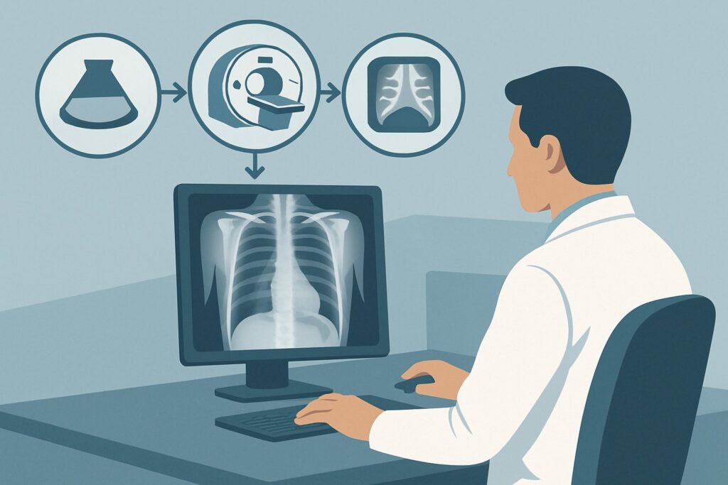
For a patient with a persistent chronic cough despite initial treatment, the ACR designates a standard posteroanterior (PA) and lateral Radiography chest as Usually Appropriate. This recommendation is based on an optimal balance of diagnostic yield, safety, and resource utilization for this specific clinical question.
The chest radiograph serves as an excellent first-line screening tool. It is widely available, inexpensive, and provides a comprehensive overview of the lungs, mediastinum, pleura, and airways. It is sufficiently sensitive to detect many of the significant pathologies in the differential, including larger nodules or masses, overt interstitial disease, significant bronchiectasis, hilar adenopathy suggestive of sarcoidosis, or consolidations from a chronic infection.
The radiation exposure from a chest radiograph is extremely low (ACR Relative Radiation Level ☢ <0.1 mSv), making it a safe initial step. This contrasts sharply with the alternatives. While CT chest without IV contrast and CT chest with IV contrast are also rated Usually Appropriate, they are generally reserved as second-line investigations. A CT provides far greater detail but comes with a substantially higher radiation dose (☢☢☢ 1-10 mSv) and cost. Starting with a chest radiograph allows clinicians to triage patients effectively, reserving the higher-dose CT for those with an abnormal radiograph or for whom clinical suspicion remains high despite a normal initial film.
Other imaging modalities are rated lower for this initial workup. For example, a Fluoroscopy biphasic esophagram is rated Usually not appropriate. While GERD is a common cause of cough, it is primarily a clinical diagnosis. An esophagram is used to evaluate for structural or motility disorders of the esophagus, which is not the primary concern in this specific scenario after empiric GERD therapy has failed to resolve the cough. The initial imaging goal is to assess the lungs, not re-evaluate for reflux.
What’s Next After the Chest Radiograph? Downstream Workflow
The results of the initial chest radiograph guide the subsequent diagnostic pathway. The workflow branches into three main possibilities:
- If the study is positive: A finding such as a lung nodule, mass, infiltrate, or signs suggestive of interstitial lung disease dictates the next step. For a suspicious nodule or mass, a CT chest with IV contrast is typically the next appropriate study to better characterize the lesion and evaluate for mediastinal involvement. If the findings suggest ILD (e.g., reticulation, ground-glass opacities), a high-resolution CT (HRCT) chest without IV contrast is the gold standard for detailed evaluation of the lung parenchyma.
- If the study is negative: A normal chest radiograph is a common and reassuring finding, but it does not end the workup for a persistent cough. This result effectively rules out gross structural disease. The focus should then shift back to the most common non-pulmonary causes (UACS, asthma, GERD), potentially with more specialized testing like spirometry with bronchoprovocation, 24-hour pH monitoring, or direct laryngoscopy. If pulmonary symptoms persist or there is a strong clinical suspicion of a condition not visible on plain film (e.g., subtle bronchiectasis or early ILD), an HRCT may be considered.
- If the study is indeterminate: Findings like a small, non-specific opacity or borderline hilar prominence can be challenging. The next step depends on the specific finding and the patient’s risk profile. This may involve a short-term follow-up radiograph to assess for stability, or proceeding directly to a CT scan for definitive characterization.
Pitfalls to Avoid (and When to Get Help)
Navigating the workup for persistent chronic cough requires careful consideration to avoid common diagnostic missteps.
A primary pitfall is prematurely ending the workup after a normal chest radiograph. A normal film is valuable for ruling out major pathology, but it does not exclude conditions like mild bronchiectasis, early ILD, or non-asthmatic eosinophilic bronchitis. If the cough persists, the investigation must continue.
Another error is ordering an advanced imaging study too early. Jumping directly to a CT scan without a preceding chest radiograph exposes the patient to unnecessary radiation and cost when a simpler test could have provided the necessary information or confirmed normalcy.
Be cautious of overlooking non-pulmonary causes. After a negative chest radiograph, it is essential to rigorously re-evaluate for UACS, GERD, and asthma-variant cough, as these remain the most frequent culprits.
If the diagnosis remains elusive despite a thorough workup including a normal chest radiograph and re-evaluation of common causes, escalation to a pulmonologist for further specialized testing is the appropriate next step.
Related ACR Topics and Tools
This article focuses on one specific clinical scenario. For a comprehensive overview of imaging for all chronic cough presentations and to explore related diagnostic challenges, the following resources are valuable.
- For breadth across all scenarios in Chronic Cough, see our parent guide: Chronic Cough: ACR Appropriateness Decoded.
- To look up appropriateness ratings for adjacent clinical scenarios, use the ACR Appropriateness Criteria Lookup.
- For detailed procedural techniques on recommended studies, consult the Imaging Protocol Library.
- To discuss cumulative radiation exposure with patients, the Radiation Dose Calculator can help frame the conversation.
Frequently Asked Questions
Why is a CT scan also rated ‘Usually Appropriate’ if a chest radiograph is the first recommended step?
Both chest radiography and CT are rated ‘Usually Appropriate’ because both can be valid initial choices depending on context. However, the ACR narrative and standard clinical practice strongly favor the chest radiograph as the *first* step due to its lower radiation dose, lower cost, and high utility as a screening tool. A CT scan is often reserved for cases where the radiograph is abnormal or when clinical suspicion for conditions like bronchiectasis or interstitial lung disease is very high despite a normal radiograph.
If the chest radiograph is normal, is a CT scan ever the right next step?
Yes. If a patient’s cough persists and is severe, and a thorough re-evaluation for non-pulmonary causes (like GERD, asthma, UACS) is unrevealing, a high-resolution CT (HRCT) of the chest may be warranted. An HRCT can detect subtle abnormalities missed on a plain radiograph, such as early interstitial lung disease or mild bronchiectasis.
Does this guidance change if the patient has a history of smoking?
Yes, significantly. A history of smoking, especially a heavy or long-term history, places the patient in a higher-risk category for lung cancer. This triggers a different ACR clinical variant (‘Chronic cough lasting more than 8 weeks. Increased risk for lung cancer. Initial imaging.’) where a non-contrast CT of the chest is often considered the most appropriate initial imaging study to screen for malignancy.
What if the patient’s cough started after a viral infection?
A post-viral or post-infectious cough is very common and can last for 3 to 8 weeks. However, once a cough persists beyond 8 weeks, it is classified as chronic. While its origin may have been post-infectious, the persistence warrants the same workup described here, starting with a chest radiograph to rule out underlying structural abnormalities that may have been unmasked or caused by the infection.
Should I order a CT of the sinuses for this patient?
A CT of the sinuses may be appropriate, but it addresses a different part of the differential. If you suspect chronic sinusitis as the cause of upper airway cough syndrome (UACS) based on clinical signs (e.g., facial pressure, purulent nasal discharge), a non-contrast CT of the maxillofacial sinuses is a reasonable test. However, it is complementary to, not a replacement for, the initial chest radiograph, which is intended to evaluate for intrathoracic causes of the cough.
Reviewed by Pouyan Golshani, MD, Interventional Radiologist — May 29, 2026