What Imaging Is Best for AxSpA Follow-Up? An ACR-Guided Workflow
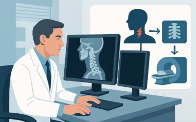
A 38-year-old patient with known ankylosing spondylitis returns to your rheumatology clinic for a 6-month follow-up after starting a TNF inhibitor. They report significant improvement in their inflammatory back pain and morning stiffness, but you need to objectively assess for structural disease progression. Deciding on the right imaging study involves balancing the need to detect subtle bony changes against the long-term risks of cumulative radiation exposure in a patient who will require lifelong monitoring. This article provides a detailed workflow for this specific clinical decision. For this scenario—monitoring a patient with known axial spondyloarthritis—the American College of Radiology (ACR) Appropriateness Criteria rate `Radiography sacroiliac joints` as Usually appropriate.
Who Fits This Clinical Scenario for Axial Spondyloarthritis Follow-Up?
This guidance applies specifically to patients with an established diagnosis of axial spondyloarthritis (axSpA). This includes both non-radiographic axSpA and the more advanced form, ankylosing spondylitis (AS). The clinical trigger for imaging is the need for longitudinal monitoring, either to assess the efficacy of a new treatment in halting structural damage or to investigate suspected disease progression during a period of clinical change or observation. The goal is to evaluate for chronic, structural changes over time, not to make an initial diagnosis.
This workflow is not intended for:
- Patients with suspected inflammatory back pain who do not yet have a diagnosis. This presentation falls under the initial imaging scenario, which typically starts with radiographs and may proceed to MRI if radiographs are negative.
- Patients with known axSpA and ankylosis who present with acute, severe pain after minimal or no trauma. This is a high-risk situation for an occult spinal fracture and requires a different imaging pathway, often involving CT.
- Patients with suspected axSpA whose initial radiographs and MRI of the sacroiliac joints were both negative. This scenario involves a different decision tree focused on alternative diagnoses or reassessing clinical criteria.
Correctly identifying the clinical question—monitoring established disease—is crucial for selecting the most appropriate and resource-conscious imaging study.
What Are You Assessing with Follow-Up Imaging in Known AxSpA?
In this scenario, the imaging study is not meant to discover a new diagnosis but to characterize the status of a known one. The key questions you are trying to answer relate to the disease’s structural impact over time.
Structural Disease Progression: This is the primary endpoint. The radiologist is looking for evidence of new or worsening chronic changes that signify ongoing disease activity despite treatment. In the sacroiliac (SI) joints, this includes new erosions, increased subchondral sclerosis, or progressive joint space narrowing and ankylosis (fusion). In the spine, the hallmark of progression is the formation or growth of syndesmophytes—bony bridges that can eventually lead to a “bamboo spine.” Detecting these changes is critical, as it may signal that the current therapeutic regimen is insufficient to halt long-term joint damage.
Stable Disease or Favorable Treatment Response: The ideal finding is the absence of any new structural changes compared to prior imaging studies. This provides objective evidence that the current treatment is effectively controlling the disease process, justifying its continuation. It is the radiographic correlate to a good clinical response.
Superimposed Degenerative Changes: Patients with long-standing inflammatory arthritis are not immune to common age-related degenerative changes, such as disc disease or facet arthropathy. While radiographs are primarily for assessing the inflammatory process, they can also help identify significant degenerative findings that may be contributing to the patient’s symptoms, potentially complicating the clinical picture.
Why Are Radiographs the Recommended Study for Monitoring AxSpA Progression?
For the routine follow-up of known axial spondyloarthritis, the ACR identifies both `Radiography sacroiliac joints` and `Radiography sacroiliac joints and spine area of interest` as Usually appropriate. This recommendation is based on a careful balance of diagnostic utility, safety, and accessibility.
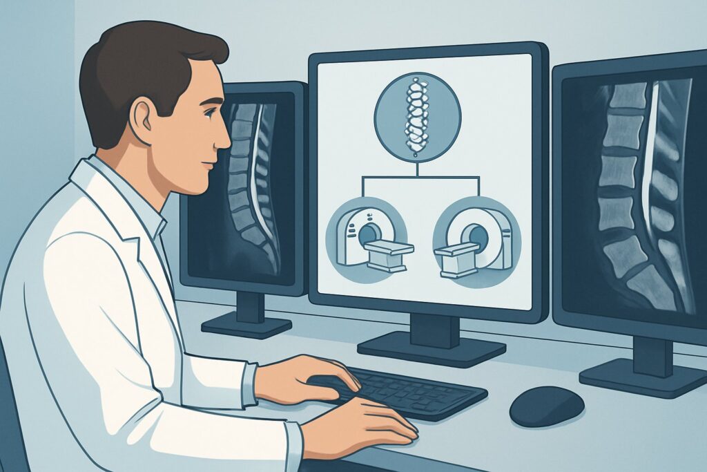
Radiography is the established gold standard for visualizing and quantifying chronic, structural bone changes. It provides excellent spatial resolution for detecting sclerosis, erosions, and the new bone formation (syndesmophytes and ankylosis) that define long-term damage in axSpA. Perhaps most importantly, radiographs are ideal for serial comparison. When current images are compared to a baseline study from one or more years prior, even subtle progression can be reliably detected. This modality is also widely available, relatively inexpensive, and quick to perform.
The ACR rates several alternative studies as less appropriate for this specific task:
- MRI of the sacroiliac joints (May be appropriate): Magnetic Resonance Imaging is superior for detecting active inflammation (bone marrow edema) and early erosions. However, it is not the primary tool for assessing the slow, chronic structural changes like sclerosis and bony fusion that are the focus of long-term monitoring. While an MRI may be indicated if the clinical question shifts to assessing acute inflammation, it is not the standard for routine structural follow-up due to higher cost and less utility for tracking syndesmophyte growth.
- CT of the sacroiliac joints (Usually not appropriate): Computed Tomography offers exceptional bone detail, but its utility for routine monitoring is severely limited by its radiation dose. A CT of the pelvis delivers a dose rated as ☢☢☢ (1-10 mSv), significantly higher than a radiograph’s ☢☢ (0.1-1 mSv). Since axSpA patients are often young and may require imaging over many decades, minimizing cumulative radiation exposure is a critical safety consideration. CT is therefore reserved for specific problems, such as evaluating a suspected fracture in an ankylosed spine.
A key ordering pearl is to ensure the radiology department has access to all relevant prior imaging studies. The comparative reading is far more valuable than a single, isolated report.
What’s the Next Step After Follow-Up Radiographs in AxSpA?
The results of follow-up radiography directly influence the downstream clinical workflow and treatment decisions. The decision tree branches based on whether structural progression is identified.
If Radiographs Show Progression: The identification of new or worsening erosions, sclerosis, or syndesmophyte formation is objective evidence of ongoing disease activity. This finding indicates that the current therapy is not adequately controlling the structural damage, even if the patient reports symptomatic improvement. The next step is a clinical re-evaluation and a discussion with the patient about escalating or changing their treatment. This may involve switching to a different class of biologic agent or adding other disease-modifying therapies.
If Radiographs Are Stable: No change from prior studies is the desired outcome. It suggests the current treatment plan is effective at halting structural progression. In this case, the typical next step is to continue the current therapy and maintain clinical monitoring. Per guidelines from groups like the Assessment of SpondyloArthritis international Society (ASAS), repeat surveillance imaging is generally not recommended more frequently than every two years in a clinically stable patient.
If Radiographs Are Stable but Symptoms Worsen: This is a common and important clinical divergence. A patient may report increased pain and stiffness despite no new changes on their radiographs. This discordance suggests that the symptoms may be driven by active inflammation (bone marrow edema), which is invisible on radiographs. This is the primary scenario where a follow-up `MRI sacroiliac joints without IV contrast` (rated May be appropriate) is warranted. Detecting active inflammation on MRI can justify a change in therapy even in the absence of new structural damage.
Common Pitfalls to Avoid in AxSpA Follow-Up Imaging
Navigating the long-term management of axial spondyloarthritis requires avoiding several common imaging-related pitfalls.
- Imaging Too Frequently: Ordering routine annual radiographs in a patient who is clinically stable and responding well to therapy provides little additional information and results in unnecessary radiation exposure. Adhere to the general principle of imaging no more than every two years unless there is a clear clinical indication.
- Forgetting to Provide Priors: The single most common error that diminishes the value of follow-up imaging is failing to make prior studies available for comparison. A radiologist’s ability to detect subtle interval change is entirely dependent on having a baseline.
- Mismatching Modality and Question: Do not order a radiograph when the primary question is about active inflammation, as it is a low-yield study for that purpose. Conversely, avoid ordering serial MRIs to track slow bony fusion when a simple radiograph is sufficient, more cost-effective, and avoids potential overuse of resources.
If a patient with an advanced, fused spine experiences a sudden increase in focal pain after even minor trauma, maintain a high suspicion for fracture. This represents a clinical emergency, and the workflow should escalate immediately to definitive imaging, which may require a CT scan.
Related ACR Topics and Tools
For a comprehensive overview of all clinical scenarios related to this topic, and for tools to help with study selection and patient communication, the following resources are available:
- For breadth across all scenarios in Inflammatory Back Pain: Known or Suspected Axial Spondyloarthropathy, see our parent guide: Inflammatory Back Pain: Known or Suspected Axial Spondyloarthropathy: ACR Appropriateness Decoded.
- To look up other clinical presentations, consult the full Imaging Appropriateness Selector tool.
- For detailed procedural techniques, see the Imaging Protocol Library.
- To help discuss radiation exposure with patients, use the Radiation Dose Calculator.
Frequently Asked Questions
How often should I order follow-up radiographs for a patient with stable axSpA?
For a patient who is clinically stable and responding well to therapy, guidelines generally recommend against imaging more frequently than every two years. More frequent imaging in the absence of a clinical change adds unnecessary radiation exposure with little chance of altering management.
My patient’s radiographs are unchanged, but their pain and stiffness are worse. What should I do?
This is a classic indication to consider an MRI. Worsening symptoms despite stable radiographs suggest active inflammation (bone marrow edema), which is not visible on x-ray. An MRI of the sacroiliac joints and/or spine without contrast is rated ‘May be appropriate’ in this scenario to detect active inflammation that could justify a change in therapy.
Why is CT rated ‘Usually not appropriate’ for routine follow-up of axSpA?
The primary reason is radiation dose. CT delivers a significantly higher dose of radiation than radiography. Since patients with axial spondyloarthritis are often young and require monitoring over their lifetime, minimizing cumulative radiation exposure is a critical safety priority. CT is therefore reserved for specific indications where its superior detail is necessary, such as a suspected fracture in an ankylosed spine.
Should I order imaging of the spine in addition to the sacroiliac joints?
Yes, this is often necessary. The ACR rates ‘Radiography sacroiliac joints and spine area of interest’ as ‘Usually appropriate.’ Disease progression, particularly the formation of syndesmophytes, can occur in the thoracic or lumbar spine even when the sacroiliac joints appear stable. The decision to image the spine should be based on the patient’s symptoms and areas of clinical concern.
Is there a role for ultrasound in monitoring axSpA?
For this specific scenario, the ACR rates ‘US sacroiliac joints’ as ‘Usually not appropriate.’ While ultrasound can be useful for evaluating peripheral enthesitis in spondyloarthritis, it is not a reliable or validated method for assessing the deep structures of the sacroiliac joints or spine for disease progression.
Reviewed by Pouyan Golshani, MD, Interventional Radiologist — May 26, 2026