What Imaging Is Best for Suspected Biceps Tendon Pain After Normal X-Rays?
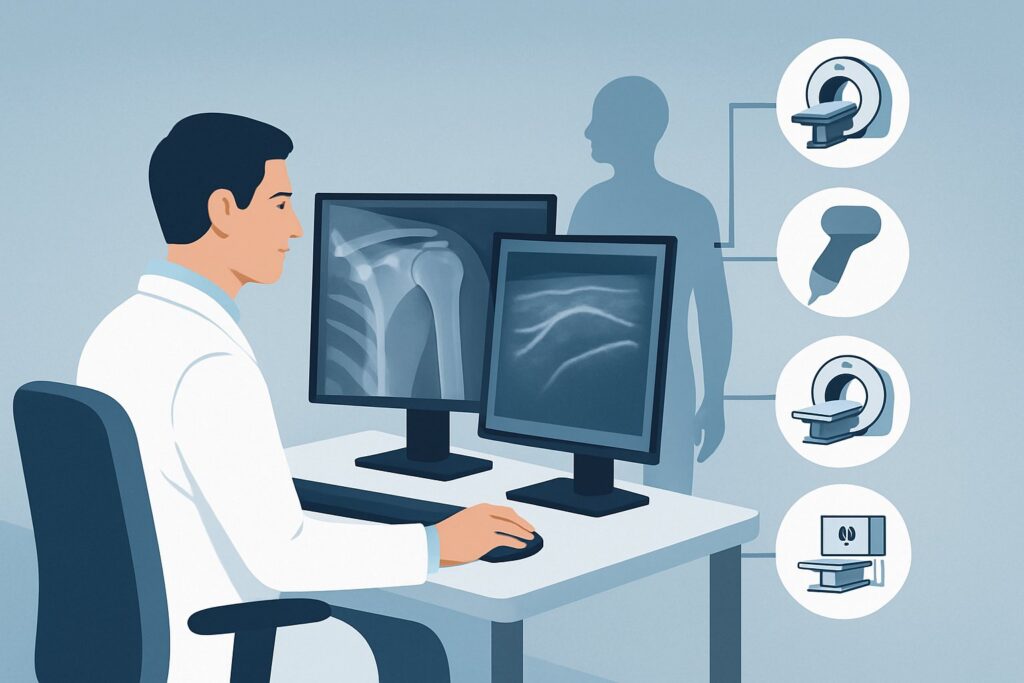
A 48-year-old patient presents with four months of nagging anterior shoulder pain, localized directly over the bicipital groove. The pain worsens with overhead activities and forearm supination, and initial radiographs were unremarkable. Your clinical suspicion is high for an abnormality of the long head of the biceps (LHB) tendon, but you need to confirm the diagnosis and assess its severity to guide treatment. What is the most appropriate next imaging study to order? This article provides a detailed clinical workflow for this specific scenario, grounded in the American College of Radiology (ACR) Appropriateness Criteria. For this presentation, shoulder ultrasound (US) is rated `Usually Appropriate` and is often the most effective initial advanced imaging test.
Who Fits This Clinical Scenario for Suspected Biceps Tendon Abnormality?
This guidance applies to a specific patient population: adults with chronic shoulder pain (lasting several weeks or more) where the clinical examination strongly suggests biceps tendon pathology. Key indicators include tenderness to palpation over the bicipital groove and positive provocative tests like Speed’s or Yergason’s. A crucial prerequisite is that initial radiographs of the shoulder have already been performed and are either normal or inconclusive, effectively ruling out significant osteoarthritis, fracture, or calcific tendinopathy as the primary pain generator.
This workflow is distinct from other common shoulder pain scenarios. It is essential to differentiate this presentation from:
- Suspected Rotator Cuff Disorders: If the patient’s primary complaint is weakness with abduction or external rotation, and pain is located more superolaterally, the workup should follow the pathway for rotator cuff pathology.
- Suspected Labral Pathology or Instability: If the history includes mechanical symptoms like clicking, catching, or a feeling of the shoulder “giving way,” especially in a younger patient, the focus shifts toward evaluating the glenoid labrum and capsule.
- Suspected Adhesive Capsulitis: If the patient presents with a global, profound loss of both active and passive range of motion in multiple planes, “frozen shoulder” is the more likely diagnosis, which has its own imaging considerations.
This article is exclusively for the patient whose history and exam point squarely at the biceps tendon as the source of their chronic pain after negative initial radiographs.
What Diagnoses Are You Working Up in This Scenario?
When you suspect a biceps tendon abnormality, you are evaluating a spectrum of conditions primarily affecting the long head of the biceps tendon as it travels through the bicipital groove and into the glenohumeral joint.
Biceps Tendinosis and Tenosynovitis
This is the most common diagnosis in this category. Tendinosis refers to the chronic, degenerative thickening and architectural disruption of the tendon itself, often from overuse. Tenosynovitis is an inflammatory process involving fluid and thickening of the tendon sheath that surrounds the LHB tendon. The two conditions frequently coexist and are a primary source of anterior shoulder pain.
Long Head of Biceps (LHB) Tendon Tear
Tears can be partial-thickness, involving only a portion of the tendon’s fibers, or full-thickness, resulting in a complete rupture. While a full-thickness rupture can sometimes produce a characteristic “Popeye” deformity, partial-thickness tears are often more subtle clinically and can be a significant cause of persistent pain and dysfunction.
LHB Tendon Instability (Subluxation or Dislocation)
This occurs when the LHB tendon moves out of its normal position within the bicipital groove. Medial subluxation or dislocation is almost always associated with an injury to the biceps pulley system, most critically a tear of the adjacent subscapularis tendon. Identifying this instability is crucial, as it changes the surgical management if an operation is considered.
Associated Subscapularis Tendon Tear
Because the subscapularis tendon and transverse humeral ligament form the primary restraint keeping the LHB tendon in the groove, pathology of the two is tightly linked. An imaging study in this scenario must also carefully evaluate the integrity of the subscapularis tendon, as an isolated biceps diagnosis might miss the root cause.
Why Is Shoulder Ultrasound the Recommended Study for This Presentation?
For a patient with suspected biceps tendon pathology and normal radiographs, both shoulder ultrasound (US) and shoulder MRI without contrast are rated `Usually Appropriate` by the ACR. However, ultrasound is often the superior first choice due to its unique diagnostic capabilities for this specific clinical question.
The primary advantage of ultrasound is its ability to perform dynamic imaging. The operator can actively move the patient’s shoulder during the scan, directly visualizing the LHB tendon to see if it subluxates or dislocates from the bicipital groove. This real-time assessment is invaluable for diagnosing instability, a finding that is static and can be missed on an MRI. Furthermore, US offers excellent spatial resolution for evaluating tendon fiber architecture, making it highly sensitive for detecting tendinosis, fluid in the tendon sheath (tenosynovitis), and partial or full-thickness tears.
Other imaging options are rated lower for specific reasons:
- MRI Shoulder without IV contrast is also `Usually Appropriate` and provides excellent visualization of the tendon and surrounding soft tissues, including the rotator cuff and labrum. It is a reasonable alternative, particularly if there is a broader differential diagnosis. However, it is static, more expensive, and less accessible than ultrasound.
- MR Arthrography Shoulder, while also `Usually Appropriate`, involves an intra-articular injection of contrast. This is more invasive than necessary when the primary question is about the extra-articular portion of the biceps tendon. It is better reserved for when intra-articular pathology, like a labral tear, is the primary concern.
- CT Arthrography Shoulder is rated `May be appropriate`. While it can show the tendon, it exposes the patient to significant ionizing radiation (ACR RRL® ☢☢☢☢, 10-30 mSv) and is less effective at evaluating tendon fiber detail compared to US or MRI.
In summary, ultrasound provides a non-invasive, radiation-free, and cost-effective method to directly answer the key clinical questions about biceps tendon integrity and stability.
What’s Next After Shoulder Ultrasound? Downstream Workflow
The results of the shoulder ultrasound will guide the subsequent steps in patient management. The decision tree typically branches based on whether the findings confirm the clinical suspicion.
If the US is positive for biceps tendinopathy or a tear:
A definitive diagnosis allows for targeted treatment. For tendinosis, tenosynovitis, or a low-grade partial tear, the next step is typically conservative management. This may include physical therapy focused on eccentric strengthening, activity modification, and anti-inflammatory medications. If symptoms persist, a referral to a sports medicine physician, physiatrist, or orthopedic surgeon for consideration of an ultrasound-guided corticosteroid injection may be appropriate. For high-grade partial or full-thickness tears, or for instability, a surgical consultation is warranted.
If the US is negative but clinical suspicion remains high:
A negative or equivocal ultrasound in a patient with persistent, classic symptoms of biceps pathology should prompt consideration of further imaging. The most logical next step is an MRI of the shoulder without contrast. This modality can provide a more global assessment of the shoulder, including the intra-articular portion of the biceps tendon, the rotator cuff, and the glenoid labrum, potentially identifying an alternative pain generator that was not apparent on the focused ultrasound exam.
If the US shows biceps instability with a subscapularis tear:
This finding significantly changes the management plan. The patient should be referred to an orthopedic surgeon, as this combined injury pattern often requires surgical repair to restore normal shoulder mechanics and resolve the instability.
Pitfalls to Avoid (and When to Get Help)
When working up suspected biceps tendon pathology, be mindful of these common pitfalls:
- Failing to request dynamic imaging: The key advantage of ultrasound is its dynamic capability. Ensure the performing sonographer or radiologist assesses for tendon subluxation during internal and external rotation of the arm.
- Attributing all anterior pain to the biceps: Remember that subscapularis tears, anterior labral pathology, and even acromioclavicular joint arthritis can present with anterior shoulder pain. Maintain a broad differential if the imaging is negative.
- Overlooking the intra-articular portion: A standard ultrasound may not fully visualize the biceps anchor at the superior labrum. If symptoms persist despite a normal extra-articular US, the problem may be inside the joint.
- Ignoring associated findings: A biceps tendon abnormality rarely occurs in isolation. Pay close attention to the report’s comments on the subscapularis and supraspinatus tendons, as these are frequently involved.
If a patient has persistent mechanical symptoms (locking, catching) or fails to improve with conservative treatment despite a “normal” ultrasound, escalate care by ordering an MRI or referring to an orthopedic specialist.
Related ACR Topics and Tools
For a comprehensive overview of imaging for all chronic shoulder pain presentations, from rotator cuff tears to osteoarthritis, please see our parent guide. For tools to help you apply these criteria in your practice, explore the resources below.
- For breadth across all scenarios in Chronic Shoulder Pain, see our parent guide: Chronic Shoulder Pain: ACR Appropriateness Decoded.
- ACR Appropriateness Criteria Lookup — for adjacent scenarios
- Imaging Protocol Library — for technique on the recommended study
- Radiation Dose Calculator — for cumulative dose conversations
Frequently Asked Questions
Why not just order an MRI for every patient with suspected biceps pain?
While MRI is also rated ‘Usually Appropriate,’ shoulder ultrasound is often preferred as the initial advanced study for suspected biceps pathology. Ultrasound is less expensive, more readily available, and offers unique dynamic imaging capabilities to assess for tendon instability (subluxation), which a static MRI cannot. It is a more targeted and efficient first step for this specific clinical question.
What if the ultrasound is negative but my patient still has classic biceps tendon pain?
If there is a strong clinical suspicion for biceps pathology despite a negative ultrasound, the next appropriate step is to order an MRI of the shoulder without contrast. The ultrasound may have missed a very subtle partial tear, or the pain generator may be intra-articular (e.g., at the biceps anchor or superior labrum), which is better visualized by MRI.
Does an ultrasound-guided corticosteroid injection have a role here?
Yes. An image-guided injection is rated ‘Usually Appropriate’ by the ACR and can serve both diagnostic and therapeutic purposes. If injecting anesthetic and corticosteroid into the biceps tendon sheath provides significant pain relief, it helps confirm the diagnosis. This is often considered after a diagnostic ultrasound has been performed and conservative measures have failed.
Is MR Arthrography ever needed for a biceps tendon issue?
MR Arthrography is generally not the first choice when the primary suspicion is an issue with the extra-articular biceps tendon. However, it becomes the preferred study if you suspect a concurrent intra-articular problem, such as a SLAP (Superior Labrum Anterior to Posterior) tear, which involves the biceps anchor complex inside the joint.
My patient’s radiograph report mentioned a ‘bicipital groove spur.’ Is that significant?
Bony spurs in or around the bicipital groove can be a source of mechanical irritation and may contribute to biceps tendinopathy or even tearing. While the radiograph identifies the spur, an ultrasound or MRI is still necessary to evaluate the condition of the tendon itself and determine if it is inflamed, thickened, or torn as a result.
Reviewed by Pouyan Golshani, MD, Interventional Radiologist — May 29, 2026