What Imaging Should You Order for Suspected Neurodegeneration with Brain Iron Accumulation?

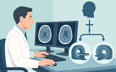
A young adult presents to your neurology clinic with a frustrating, years-long history of progressive dystonia and parkinsonism. The symptoms began subtly in adolescence and have now advanced to include cognitive slowing and dysarthria. Standard workups have been unrevealing, but the combination of early-onset movement disorder and cognitive decline raises suspicion for a genetic etiology, specifically a form of neurodegeneration with brain iron accumulation (NBIA). You need to order imaging to confirm the presence of iron deposition and narrow the differential. This article details the ACR-guided workflow for this specific clinical question. For the initial imaging of suspected NBIA, an MRI of the head without IV contrast is rated as Usually Appropriate.
Who Fits the Scenario for Suspected Neurodegeneration with Brain Iron Accumulation?
This clinical workflow is designed for patients, typically children or young adults, presenting with a progressive and complex movement disorder. The core features prompting suspicion of NBIA include a combination of extrapyramidal symptoms such as dystonia, parkinsonism, and choreoathetosis. These are often accompanied by other neurologic signs like spasticity, pyramidal tract signs, optic atrophy, retinal degeneration, or cognitive decline.
It is crucial to distinguish this specific presentation from similar, but distinct, clinical scenarios that require different imaging workups:
- Exclusion 1: Classic Parkinsonian Syndromes. An older adult presenting with the classic triad of resting tremor, rigidity, and bradykinesia, without the atypical features seen in NBIA, fits the “Parkinsonian syndromes” scenario.
- Exclusion 2: Isolated Chorea. A patient presenting with pure chorea and a strong family history of a similar disorder is better evaluated under the “Chorea; suspected Huntington disease” pathway.
- Exclusion 3: Rapidly Progressive Dementia. A patient experiencing a precipitous cognitive decline over weeks to months, with or without motor signs, aligns with the workup for “Rapidly progressive dementia; suspected Creutzfeldt-Jakob disease.”
This guide is for the specific instance where the clinical picture points toward a primary disorder of brain iron metabolism as the underlying cause of a complex, progressive neurodegenerative process.
What Diagnoses Are You Working Up with Suspected Brain Iron Accumulation?
The primary diagnostic category you are investigating is Neurodegeneration with Brain Iron Accumulation (NBIA), a group of rare, inherited neurologic disorders. Imaging plays a critical role in identifying the hallmark feature of these conditions and can often suggest a specific genetic subtype, guiding subsequent molecular testing. The differential includes several key disorders.
Pantothenate kinase-associated neurodegeneration (PKAN) is the most common form of NBIA. It is caused by mutations in the PANK2 gene and typically presents in childhood with gait impairment, dystonia, and dysarthria. The imaging findings in PKAN are often pathognomonic, making MRI a powerful diagnostic tool.
PLA2G6-associated neurodegeneration (PLAN) encompasses a spectrum of disorders, including infantile neuroaxonal dystrophy (INAD), atypical neuroaxonal dystrophy (NAD), and an adult-onset dystonia-parkinsonism. In addition to iron accumulation, cerebellar atrophy is a prominent feature that MRI can clearly delineate.
Beta-propeller protein-associated neurodegeneration (BPAN) is an X-linked dominant disorder that presents differently depending on the patient’s age. It often begins with developmental delay and seizures in childhood, followed by the onset of progressive dystonia and parkinsonism in adolescence or early adulthood. The pattern of iron deposition can be characteristic.
Aceruloplasminemia is an autosomal recessive disorder of iron metabolism that presents in adulthood. Neurologic symptoms include ataxia, dystonia, and cognitive impairment, but patients also have systemic manifestations like retinal degeneration and diabetes mellitus. Iron deposition on MRI is typically more widespread than in other NBIA forms, involving the thalamus and dentate nuclei in addition to the basal ganglia.
Why Is MRI Head without IV Contrast the Recommended Study for Suspected NBIA?
The American College of Radiology designates MRI of the head without IV contrast as “Usually Appropriate” for the initial evaluation of suspected NBIA because of its unparalleled ability to detect iron deposition and associated structural changes without exposing the patient to ionizing radiation or contrast agents.
The diagnostic strength of MRI lies in its sensitivity to the paramagnetic properties of iron. Iron accumulation causes local magnetic field distortions, which result in signal loss (darkening) on specific MRI sequences. T2-weighted images are fundamental for this evaluation, but gradient-recalled echo (GRE) and, more modernly, susceptibility-weighted imaging (SWI) sequences are exceptionally sensitive to iron and are considered essential components of the imaging protocol. These sequences can detect subtle iron deposition long before it might become apparent on other imaging modalities.
Furthermore, MRI can reveal highly specific patterns that point toward a particular NBIA subtype. The classic example is the “eye-of-the-tiger” sign seen in many patients with PKAN. This sign consists of marked T2 hypointensity (darkness) in the globus pallidus due to iron accumulation, surrounding a central region of T2 hyperintensity (brightness) thought to represent gliosis and neuronal loss. Identifying this sign can dramatically narrow the differential and streamline the diagnostic process.
Why Other Studies Are Rated Lower
Alternative imaging studies are considered less appropriate for this specific clinical question:
- CT head without IV contrast is rated “May be appropriate.” While severe, long-standing iron deposition can sometimes manifest as hyperdensity (or associated calcification) in the basal ganglia on CT, this modality is significantly less sensitive than MRI. CT is generally reserved for situations where MRI is contraindicated or unavailable. It also involves ionizing radiation (☢☢☢ 1-10 mSv for adults, ☢☢☢ 0.3-3 mSv for pediatrics), a key consideration in a typically young patient population that may require follow-up imaging.
- MRI head without and with IV contrast is also rated “May be appropriate.” However, the addition of gadolinium-based contrast is not necessary for the primary goal of identifying iron. NBIA disorders are not typically associated with inflammation or blood-brain barrier breakdown that would necessitate contrast enhancement. Omitting contrast avoids potential risks, such as nephrogenic systemic fibrosis in patients with renal impairment and concerns about gadolinium deposition, without sacrificing diagnostic information relevant to this scenario.
- FDG-PET/CT brain is rated “Usually not appropriate.” While PET can show patterns of hypometabolism, it does not directly visualize iron deposition, which is the key pathologic feature. It also involves significant radiation exposure (☢☢☢ 1-10 mSv for adults, ☢☢☢☢ 3-10 mSv for pediatrics).
What’s Next After MRI Head without IV Contrast? Downstream Workflow
The results of the initial MRI will guide the subsequent diagnostic and management steps. The workflow typically proceeds down one of three paths.
If the study is positive for iron accumulation: A positive finding, especially with a characteristic pattern like the “eye-of-the-tiger” sign, provides strong evidence for an NBIA disorder. The next step is targeted genetic testing. The specific pattern of iron deposition (e.g., globus pallidus vs. substantia nigra and globus pallidus) and other associated findings (e.g., cerebellar atrophy, cortical atrophy) can help prioritize which genes to test first, making the genetic workup more efficient. The patient should be referred to a movement disorders specialist and a genetic counselor to discuss the implications of these findings and coordinate testing.
If the study is negative: A normal MRI that shows no evidence of abnormal iron accumulation makes a classic NBIA disorder much less likely, prompting a re-evaluation of the differential diagnosis. The clinical focus may shift to other causes of young-onset parkinsonism or dystonia, such as Wilson disease, dopa-responsive dystonia, or other genetic movement disorders. Further workup might include laboratory testing (e.g., serum ceruloplasmin and copper levels) or pursuing broader genetic panels for movement disorders.
If the study is indeterminate or shows non-specific findings: Occasionally, an MRI may show subtle or atypical signal changes in the basal ganglia that are not definitive for NBIA. In these cases, the clinical correlation is paramount. The decision may be to proceed with broad panel-based genetic testing for NBIA and other look-alike disorders. In some instances, a follow-up MRI in 1-2 years may be considered to assess for progression of signal changes, which would increase suspicion for a neurodegenerative process.
Pitfalls to Avoid (and When to Get Help)
When working up suspected NBIA, be mindful of these common pitfalls:
- Inadequate MRI Protocol: Ordering a “routine” brain MRI may not include the specific sequences (like SWI or GRE) that are most sensitive for iron. It is crucial to specify “evaluation for brain iron” or “NBIA protocol” on the imaging requisition to ensure the radiologist performs the necessary sequences.
- Misinterpreting Age-Related Iron: A small amount of iron deposition in the basal ganglia can be a normal finding in older adults. Interpreting this as pathologic in the absence of a compelling clinical history is a potential error; correlation with the patient’s age and symptoms is essential.
- Overlooking Mimics: Conditions like Wilson disease, mitochondrial disorders, or even chronic liver disease can cause basal ganglia signal abnormalities. Do not anchor on NBIA without considering and ruling out key mimics.
- Delaying Genetic Counseling: Given that NBIA disorders are genetic, involving a genetic counselor early in the process is critical for managing patient and family expectations and planning for confirmatory testing.
If the clinical picture and imaging findings are discordant or confusing, escalation to a multidisciplinary team including a neuroradiologist and a neurologist specializing in movement disorders is the appropriate next step.
Related ACR Topics and Tools
This article focuses on a single clinical scenario. For a comprehensive overview of imaging for other related conditions, from parkinsonian syndromes to chorea, consult the parent topic guide. For additional resources on ordering appropriate imaging, see the tools below.
- For breadth across all scenarios in Movement Disorders and Neurodegenerative Diseases, see our parent guide: Movement Disorders and Neurodegenerative Diseases: ACR Appropriateness Decoded.
- To look up other clinical scenarios, use the ACR Appropriateness Criteria Lookup.
- To understand the technical details of the recommended study, explore the Imaging Protocol Library.
- To discuss radiation exposure with patients, especially when considering CT, use the Radiation Dose Calculator.
Frequently Asked Questions
Why is susceptibility-weighted imaging (SWI) so important for an NBIA workup?
Susceptibility-weighted imaging (SWI) is an MRI sequence that is exceptionally sensitive to substances that distort the local magnetic field, such as iron and blood products. It provides much greater contrast and conspicuity for iron deposition than standard T2-weighted images, allowing for the detection of more subtle abnormalities and a more confident diagnosis. It is considered an essential part of a modern MRI protocol for suspected NBIA.
Can MRI definitively diagnose a specific type of NBIA?
While MRI can be highly suggestive of a specific NBIA subtype, it is not a definitive diagnosis on its own. The classic ‘eye-of-the-tiger’ sign is strongly associated with PKAN, but it has been reported in other conditions. Ultimately, the diagnosis must be confirmed with molecular genetic testing. The MRI findings are crucial for guiding which genetic tests to order first.
Is there a role for follow-up imaging if the initial MRI is positive?
In most cases, once a diagnosis of NBIA is confirmed by genetic testing, routine follow-up imaging is not necessary for clinical management. Imaging is primarily a diagnostic tool in this context. However, follow-up scans may be performed as part of a research study to track disease progression or in atypical cases where the diagnosis remains uncertain and monitoring for changes over time could be informative.
What should I do if my patient has a contraindication to MRI, like a non-compatible pacemaker?
If MRI is absolutely contraindicated, a CT head without IV contrast is the next best option, rated as ‘May be appropriate’ by the ACR. You must be aware that CT is much less sensitive for detecting iron, and a negative CT scan does not rule out NBIA. The report should be interpreted with caution, and the decision to proceed with genetic testing would have to be based more heavily on the clinical suspicion alone.
Does the absence of the ‘eye-of-the-tiger’ sign rule out PKAN?
No. While the ‘eye-of-the-tiger’ sign is a classic finding in pantothenate kinase-associated neurodegeneration (PKAN), it is not present in all patients. Its absence, especially early in the disease course, does not exclude the diagnosis. Other patterns of globus pallidus iron accumulation can be seen, and if clinical suspicion for PKAN is high, genetic testing should still be considered.
Reviewed by Pouyan Golshani, MD, Interventional Radiologist — May 29, 2026