What Is the Best Imaging for a Soft Tissue Mass When MRI Is Contraindicated?
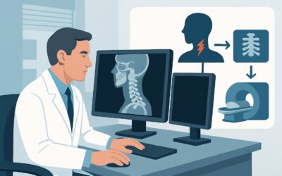
A 58-year-old patient with a recently placed cardiac pacemaker presents to your clinic with a palpable, deep soft tissue mass in his thigh that has been slowly enlarging. The initial workup included a radiograph, which was negative for osseous abnormality, and a noncontrast enhanced ultrasound, which confirmed a solid, indeterminate 5 cm mass without definitive features. Magnetic Resonance Imaging (MRI), the typical next step for soft tissue characterization, is contraindicated due to his new device. You are now faced with a critical decision: what is the most appropriate next imaging study to evaluate this mass and guide management?
This article provides a detailed clinical workflow for this specific scenario, grounded in the American College of Radiology (ACR) Appropriateness Criteria. For this presentation, the ACR designates CT of the area of interest with IV contrast as Usually Appropriate, providing a crucial alternative when MRI is not an option.
Who Fits This Clinical Scenario?
This guidance is specifically for patients presenting with a soft tissue mass where the initial, foundational imaging has been performed but remains inconclusive, and the gold-standard study for soft tissue is unavailable.
Inclusion criteria for this workflow:
- The patient has a clinically identified soft tissue mass.
- A radiograph of the area has been performed and is nondiagnostic (e.g., shows no calcification, osseous erosion, or foreign body to explain the mass).
- A noncontrast enhanced ultrasound has been performed and is also nondiagnostic, typically showing a solid or complex mass that cannot be definitively characterized as benign.
- The patient has an absolute or strong relative contraindication to MRI, such as an incompatible cardiac pacemaker or defibrillator, certain cochlear implants, retained metallic foreign bodies in a critical location, or severe, unmanageable claustrophobia.
It is critical to distinguish this situation from related but distinct clinical presentations. This workflow does not apply if:
- The patient is eligible for MRI. If MRI is an option, it is the preferred next step after nondiagnostic radiograph and ultrasound. This represents a different branch in the ACR guidelines.
- No initial imaging has been performed. For a palpable deep soft tissue mass, the initial imaging choice itself is a separate clinical question.
- The mass is clearly superficial. A small, mobile, superficial mass may have a different workup pathway, often starting and ending with ultrasound.
What Diagnoses Are You Working Up in This Scenario?
When initial imaging is indeterminate and MRI is off the table, the primary goal of the next study is to identify features that can differentiate between benign and malignant processes, with the chief concern being a soft tissue sarcoma. The differential diagnosis guides the choice of imaging and the interpretation of its findings.
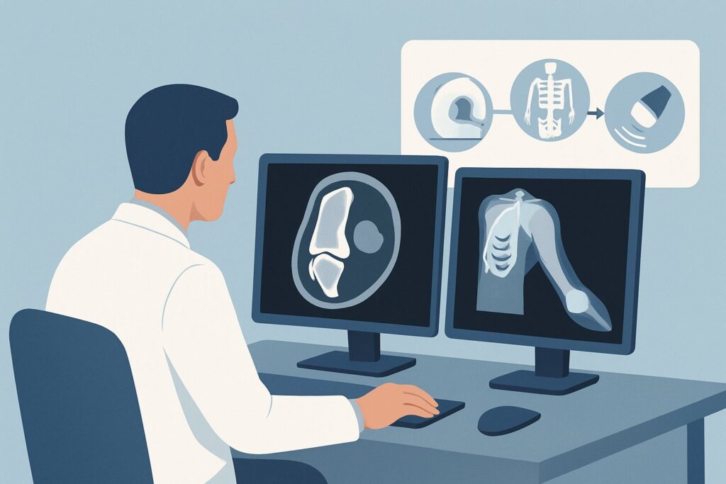
Soft Tissue Sarcoma: This is the most consequential diagnosis to exclude. These malignant tumors can arise from mesenchymal tissues like fat, muscle, and nerves. On imaging, the goal is to identify features suggestive of malignancy, such as large size (>5 cm), deep location, heterogeneous composition, areas of necrosis, and invasion of adjacent structures. Contrast-enhanced CT is valuable for assessing these features, particularly tumor vascularity and necrosis.
Benign Non-Lipomatous Tumors: This category includes a wide range of entities such as schwannomas, neurofibromas, and desmoid tumors. While benign, some can be locally aggressive or cause significant symptoms. Contrast-enhanced CT can help characterize their vascular supply and relationship to surrounding neurovascular bundles and fascial planes, which is critical for surgical planning.
Atypical Lipomatous Tumor / Well-Differentiated Liposarcoma: While ultrasound can often identify simple lipomas, more complex fatty masses may be indeterminate. Contrast-enhanced CT can reveal non-fatty components, thick septa, or nodular enhancement that would raise suspicion for a well-differentiated liposarcoma, which requires oncologic management.
Inflammatory or Infectious Processes: An abscess or organized hematoma can mimic a solid tumor on noncontrast ultrasound. The enhancement pattern on a contrast-enhanced CT—typically peripheral or rim-enhancement for an abscess—can often provide a definitive diagnosis and guide percutaneous drainage rather than surgical excision.
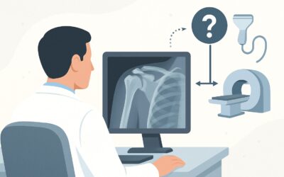
Why Is CT with IV Contrast the Recommended Study for This Presentation?
When MRI is contraindicated, contrast-enhanced Computed Tomography (CT) becomes the primary problem-solving tool for characterizing an indeterminate soft tissue mass. The ACR rates CT of the area of interest with IV contrast as Usually Appropriate because it provides the most diagnostic information among the available non-MRI modalities.
The rationale is rooted in CT’s ability to assess key features of a mass that help stratify its risk of malignancy:
- Vascularity and Enhancement: Intravenous contrast opacifies blood vessels, allowing assessment of a mass’s vascularity. Malignant tumors, particularly high-grade sarcomas, are often hypervascular and show avid, heterogeneous enhancement. In contrast, simple cysts or chronic hematomas will not enhance.
- Necrosis: The absence of enhancement within a mass is a strong indicator of necrosis, a feature commonly seen in rapidly growing malignant tumors.
- Anatomic Relationships: CT provides excellent spatial resolution, clearly defining the mass’s size, location (superficial vs. deep to fascia), and its relationship to adjacent bone, muscle compartments, and neurovascular structures. This information is vital for pre-biopsy and pre-surgical planning.
In contrast, other imaging options are rated lower for this specific scenario:
- CT area of interest without IV contrast is rated Usually Not Appropriate. Without contrast, CT offers limited soft-tissue differentiation. It is difficult to distinguish a solid tumor from muscle, or to identify vascularity and necrosis, which are the key features needed for characterization at this stage.
- Image-guided biopsy is also rated Usually Not Appropriate as the next diagnostic step. Proceeding directly to biopsy without advanced cross-sectional imaging is a significant potential pitfall. Imaging is required to characterize the lesion, identify the most viable and aggressive-appearing components to target, and plan a safe biopsy trajectory that does not contaminate uninvolved tissue compartments.
The radiation dose for a CT scan is a consideration (ACR Relative Radiation Level: Varies). The risk must be weighed against the benefit of accurately diagnosing or excluding a potentially life-threatening malignancy.
What’s Next After CT with IV Contrast? Downstream Workflow
The results of the contrast-enhanced CT will direct the subsequent steps in patient management. The workflow diverges based on whether the findings are suspicious for malignancy, clearly benign, or remain indeterminate.
- If the CT findings are suspicious for malignancy: A mass that is large, deep, and demonstrates heterogeneous enhancement or central necrosis requires urgent referral to a sarcoma specialist, typically an orthopedic or surgical oncologist. The next step is almost always a core needle biopsy, planned by the treating surgeon in conjunction with a radiologist, using the CT images to guide the approach. Further staging studies, such as a chest CT, may also be indicated.
- If the CT findings are definitively benign: If the CT characterizes the mass as a simple fluid collection (e.g., hematoma, seroma) or a classic benign entity, management may consist of clinical observation, reassurance, or symptomatic treatment. For example, a confirmed abscess would proceed to drainage.
- If the CT findings are indeterminate: In some cases, even a contrast-enhanced CT may not be able to definitively distinguish between a benign and malignant process. These cases often require a multidisciplinary discussion and will typically proceed to an image-guided core needle biopsy to obtain a tissue diagnosis. The CT provides the necessary roadmap for performing this biopsy safely and effectively.
Pitfalls to Avoid (and When to Get Help)
Navigating this clinical scenario requires careful attention to detail to avoid common errors that can delay diagnosis or compromise treatment.
- Pitfall 1: Ordering a noncontrast CT. As noted, a CT without IV contrast provides minimal additional information over ultrasound for soft tissue characterization and is rated “Usually Not Appropriate.” Always specify “with IV contrast” unless there is a contraindication like severe renal insufficiency or contrast allergy.
- Pitfall 2: Insufficient clinical history. The radiologist’s interpretation is significantly enhanced by relevant history. Always include the patient’s age, mass location, duration, rate of growth, and history of trauma or prior malignancy.
- Pitfall 3: Premature biopsy. Biopsy should follow, not precede, advanced imaging. An improperly planned biopsy can compromise a limb-sparing cancer surgery.
- Pitfall 4: Assuming an MRI contraindication is permanent. Some contraindications are relative or temporary. It is worth confirming the specific device model and its MRI compatibility, as many modern pacemakers and implants are now MRI-conditional.
If CT findings are suspicious for sarcoma or remain indeterminate after a complete non-invasive workup, this is the point to escalate care by referring the patient to a specialized center with expertise in musculoskeletal oncology.
Related ACR Topics and Tools
For a comprehensive overview of imaging recommendations across all soft tissue mass presentations, and for tools to help with study selection and patient communication, the following resources are available:
- For breadth across all scenarios in Soft Tissue Masses, see our parent guide: Soft Tissue Masses: ACR Appropriateness Decoded.
- To explore the ACR criteria for thousands of other clinical scenarios, use the ACR Appropriateness Criteria Lookup tool.
- To review technical parameters for various imaging studies, consult the Imaging Protocol Library.
- For discussions about radiation exposure with patients, the Radiation Dose Calculator can help quantify and contextualize the dose from CT scans.
Frequently Asked Questions
Why can’t I just order a biopsy after the nondiagnostic ultrasound?
The ACR rates image-guided biopsy as ‘Usually Not Appropriate’ as the immediate next step because advanced imaging (like CT in this case) is crucial for pre-biopsy planning. The CT scan helps identify the most aggressive parts of the tumor to target, avoiding necrotic areas that would yield a non-diagnostic sample. It also maps out a safe path for the needle that avoids contaminating uninvolved muscle compartments, which is critical for future limb-sparing surgery if the mass is a sarcoma.
What if my patient has a severe contrast allergy or renal failure and cannot receive IV contrast?
This creates a more challenging diagnostic dilemma. A noncontrast CT is of limited value. In this situation, a multidisciplinary discussion with radiology and potentially a sarcoma specialist is warranted. Options might include re-evaluating the MRI contraindication (as some devices are MRI-conditional), considering a contrast-enhanced ultrasound (CEUS) if available and expertise exists, or proceeding directly to a carefully planned image-guided biopsy, accepting the limitations of not having full pre-procedural characterization.
Is a PET/CT scan a good alternative to a diagnostic CT with contrast?
No, not for initial characterization. The ACR rates FDG-PET/CT as ‘Usually Not Appropriate’ for this specific scenario. While PET/CT is excellent for staging known malignancies and assessing treatment response, it has lower spatial resolution than a diagnostic CT and is not the primary tool for the initial characterization of an unknown soft tissue mass. It also involves a significantly higher radiation dose.
How do I confirm if a patient’s pacemaker or implant is truly an absolute contraindication to MRI?
It is essential to verify the specific make and model of the implanted device. Many modern cardiac devices and implants are ‘MRI-conditional,’ meaning they are safe for MRI under specific protocols. This requires checking the device card, consulting the manufacturer’s documentation, and coordinating closely with the radiology department and the patient’s cardiologist or the service that implanted the device. Never assume a device is an absolute contraindication without verification.
Does the location of the soft tissue mass (e.g., arm vs. leg vs. trunk) change this recommendation?
No, the fundamental recommendation for CT with IV contrast remains the same regardless of the location of the deep soft tissue mass. The technical parameters of the CT scan, such as the scan range and field of view, will be tailored to the specific ‘area of interest,’ but the choice of modality and the need for intravenous contrast do not change based on location.
Reviewed by Pouyan Golshani, MD, Interventional Radiologist — May 29, 2026