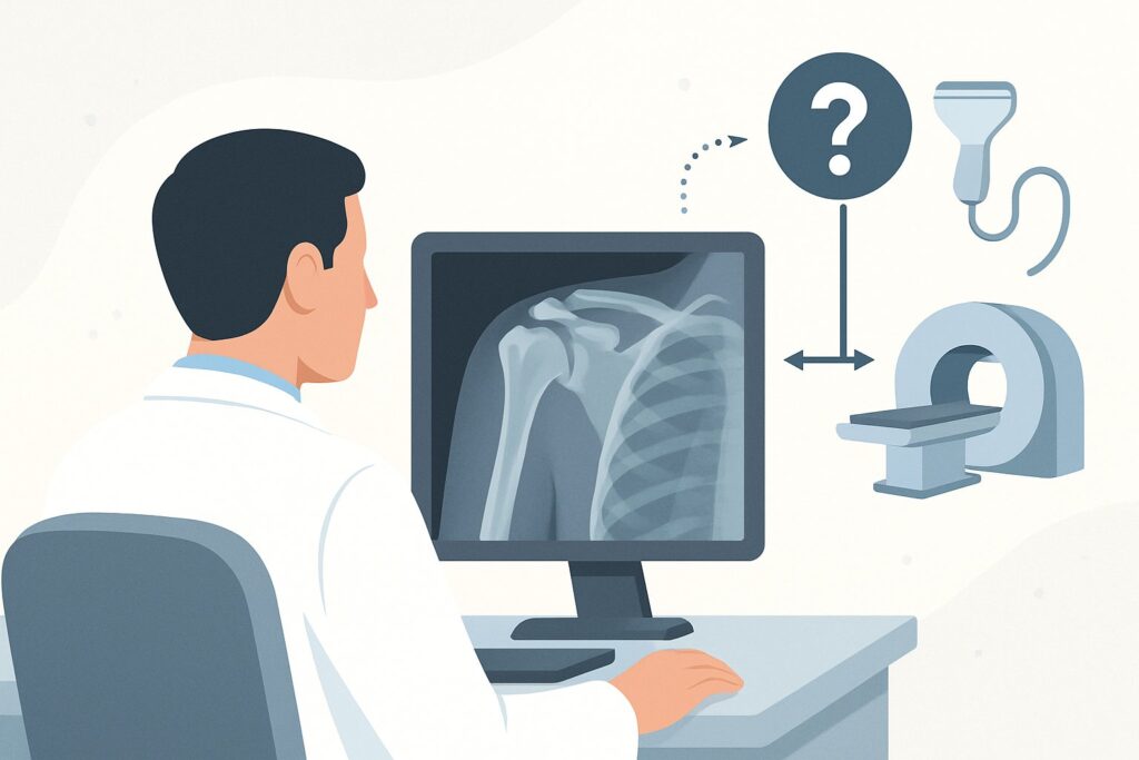
Should You Order Ultrasound or MRI for Chronic Shoulder Pain After Normal X-Rays?
A 55-year-old patient presents with six months of nagging right shoulder pain, localized over the deltoid, which worsens when he reaches overhead. His physical exam suggests impingement, and you suspect a rotator cuff disorder. You ordered initial radiographs, which came back normal, showing no fracture, significant osteoarthritis, or calcific deposits. Now you face the next decision point: which advanced imaging study will best evaluate the tendons and bursa? This article provides a detailed clinical workflow for this specific scenario, grounded in the American College of Radiology (ACR) Appropriateness Criteria, which rates shoulder ultrasound as Usually Appropriate for this indication.
Who Fits This Clinical Scenario?
This guidance is for a specific patient population: adults with chronic shoulder pain (lasting weeks to months) where the clinical suspicion points toward rotator cuff pathology or subacromial-subdeltoid bursitis. Key inclusion criteria for this workflow are:
- No history of prior shoulder surgery.
- Initial radiographs have already been performed and are either normal or inconclusive (e.g., show minor degenerative changes not sufficient to explain the symptoms).
- The primary clinical question is the integrity of the rotator cuff and the status of the adjacent bursa.
This workflow is not appropriate for patients with different primary suspicions. For instance, if the patient’s history involves significant trauma, recurrent dislocations, or a feeling of instability, the primary concern shifts to labral pathology or ligamentous injury, which follows a different diagnostic algorithm. Similarly, a patient presenting with global, profound loss of both active and passive range of motion is more likely suffering from adhesive capsulitis. If initial radiographs clearly demonstrate advanced glenohumeral osteoarthritis or large calcific deposits, the subsequent imaging choice and management plan also diverge from this pathway.
What Diagnoses Are You Working Up in This Scenario?
When initial radiographs are unrevealing in a patient with chronic shoulder pain, the differential diagnosis shifts from bone-centric problems to soft-tissue pathology. The imaging study you order next is intended to directly visualize these structures.
Rotator Cuff Tendinopathy and Tears: This is the most common diagnosis in this scenario. It represents a spectrum from inflammation and thickening of the tendons (tendinopathy) to partial-thickness tears (fraying on the articular or bursal side) to full-thickness tears. The supraspinatus tendon is most frequently involved due to its anatomical position beneath the acromion. The goal of imaging is to confirm the diagnosis, grade the severity of the tear, and assess for secondary signs like muscle atrophy, which can influence treatment decisions.
Subacromial-Subdeltoid Bursitis: The subacromial-subdeltoid bursa is a fluid-filled sac that reduces friction between the rotator cuff tendons and the overlying acromion. Inflammation, or bursitis, is a frequent cause of shoulder pain and often co-exists with rotator cuff tendinopathy. Imaging can identify bursal fluid and thickening, confirming it as a pain generator.
Biceps Tendinopathy or Instability: While the primary suspicion may be the rotator cuff, the long head of the biceps tendon is intimately related and can be a concurrent source of pain. Imaging can evaluate for tendinosis, tenosynovitis (fluid in the tendon sheath), or subluxation/dislocation of the tendon from the bicipital groove.
Why Is Shoulder Ultrasound the Recommended Study for This Presentation?
For a patient with suspected rotator cuff pathology and normal radiographs, the ACR rates three studies as Usually Appropriate: Ultrasound (US) shoulder, MRI shoulder without IV contrast, and MR arthrography shoulder. While all are highly rated, ultrasound often emerges as the preferred initial choice for several practical and clinical reasons.
Ultrasound (US) shoulder (0 mSv) is an excellent first-line modality. Its primary advantage is the ability to perform a dynamic assessment. The sonographer can visualize the rotator cuff tendons and subacromial space in real-time as the patient moves their arm, directly demonstrating impingement. US provides extremely high spatial resolution for evaluating tendon fiber architecture, making it highly sensitive and specific for detecting tendinosis and tears. It is also quick, widely available, cost-effective, and involves no ionizing radiation. It is the ideal test for patients with contraindications to MRI.
MRI shoulder without IV contrast (0 mSv) is also Usually Appropriate and provides a more global, comprehensive view of the entire shoulder joint. It offers superb soft-tissue contrast, allowing for detailed evaluation of the rotator cuff muscles and tendons, labrum, cartilage, and bone marrow. While it cannot perform a dynamic assessment, it is less operator-dependent than ultrasound and may be superior for detecting subtle partial-thickness articular-sided tears or assessing muscle atrophy and fatty infiltration, which are important for surgical planning.
In contrast, some alternative studies are rated lower for this specific clinical question. CT arthrography shoulder is rated as May be appropriate. It is an excellent alternative for evaluating rotator cuff tears in patients who cannot undergo MRI, but it is invasive (requires an intra-articular injection) and involves a significant radiation dose (☢☢☢☢ 10-30 mSv). Furthermore, MRI shoulder without and with IV contrast is rated Usually not appropriate. For isolated rotator cuff or bursal pathology, the addition of gadolinium-based contrast provides little to no additional diagnostic information over a non-contrast study and adds unnecessary risk and cost.
What’s Next After Shoulder Ultrasound? Downstream Workflow
The results of the shoulder ultrasound will guide your subsequent management steps. The workflow typically branches into one of three paths.
If the study is positive for a full-thickness rotator cuff tear: This finding often warrants a referral to an orthopedic surgeon or sports medicine specialist. The report will typically detail the size of the tear, the degree of tendon retraction, and the condition of the muscle belly (e.g., atrophy), all of which are critical for determining whether the patient is a candidate for surgical repair versus non-operative management.
If the study shows tendinopathy, a partial-thickness tear, or bursitis: These findings are often managed conservatively, at least initially. The next step is typically a structured physical therapy program focused on strengthening the periscapular muscles and rotator cuff. An image-guided corticosteroid injection into the subacromial-subdeltoid bursa, which is rated as May be appropriate by the ACR, can be considered for diagnostic and therapeutic purposes, especially if bursitis is the dominant finding.
If the study is negative or inconclusive: If the ultrasound is normal but your clinical suspicion for an intra-articular derangement remains high, the next logical step is to obtain an MRI shoulder without IV contrast. MRI can provide a more comprehensive view, potentially identifying pathology missed on ultrasound, such as a small articular-sided tear, bone marrow edema, or an unexpected diagnosis like a paralabral cyst or early avascular necrosis.
Pitfalls to Avoid (and When to Get Help)
Navigating the workup for chronic shoulder pain requires careful correlation of imaging with the clinical picture. One major pitfall is failing to recognize the high prevalence of asymptomatic rotator cuff tears, especially in older populations. An imaging finding of a tear does not automatically mean it is the source of the patient’s pain; treatment should always be guided by the physical exam and patient symptoms.
Another common issue is the operator dependency of ultrasound. The quality and accuracy of a shoulder US are highly dependent on the skill of the sonographer and the interpreting radiologist. If the results from a community imaging center seem incongruous with your clinical exam, consider repeating the study at a center with dedicated musculoskeletal ultrasound expertise.
Finally, avoid ordering an MRI with intravenous contrast for this indication. As noted by the ACR, it is “Usually not appropriate” and adds no value for evaluating the rotator cuff or bursa, while increasing cost and exposing the patient to gadolinium.
If a patient presents with red flag symptoms such as fever, unexplained weight loss, or a rapidly progressing neurologic deficit, escalate immediately for a more urgent and extensive workup beyond this routine imaging pathway.
Related ACR Topics and Tools
This article focuses on a single, common clinical scenario. For a comprehensive overview of all variants and recommendations for chronic shoulder pain, please consult our parent guide. For additional resources on imaging selection, protocols, and radiation safety, explore the tools below.
- For breadth across all scenarios in Chronic Shoulder Pain, see our parent guide: Chronic Shoulder Pain: ACR Appropriateness Decoded.
- To explore other clinical situations, use the ACR Appropriateness Criteria Lookup.
- For details on imaging techniques, visit the Imaging Protocol Library.
- To discuss radiation exposure with patients, consult the Radiation Dose Calculator.
Frequently Asked Questions
Why are both ultrasound and MRI rated ‘Usually Appropriate’ for suspected rotator cuff issues?
Both modalities have excellent diagnostic accuracy for detecting rotator cuff tears. The American College of Radiology rates them equally because the choice often depends on local expertise, cost, availability, and specific secondary questions. Ultrasound excels in dynamic assessment and tendon detail, while MRI provides a more comprehensive, global view of all joint structures. For the primary question of a rotator cuff tear, either is an excellent choice.
If MRI is an option, why would I ever start with ultrasound?
Ultrasound is often preferred as the initial advanced imaging test due to several advantages: it is typically less expensive, more widely available with shorter wait times, and involves no contraindications like pacemakers or claustrophobia. Its ability to perform a real-time dynamic evaluation to visualize impingement is a unique benefit that MRI cannot replicate.
What specific findings should I look for in the ultrasound report?
Key findings to look for include: the presence and size of any full- or partial-thickness rotator cuff tears (specifying which tendon is involved), the degree of tendon retraction for full-thickness tears, signs of tendinosis (thickening and hypoechogenicity), fluid in the subacromial-subdeltoid bursa, and any abnormalities of the long head of the biceps tendon.
When is MR arthrography a better choice than a non-contrast MRI for this scenario?
While both are rated ‘Usually Appropriate,’ MR arthrography is more invasive as it requires injecting contrast directly into the joint. It is generally reserved for cases where there is a high suspicion of a small partial-thickness tear on the articular side of the cuff or when there is a concurrent suspicion of labral pathology or instability, as the injected contrast helps delineate these structures more clearly.
My patient has a pacemaker and can’t get an MRI. What is the next best test after inconclusive radiographs?
Ultrasound is the ideal next step in this situation. It provides excellent visualization of the rotator cuff and bursa without the contraindications of MRI. If ultrasound is also inconclusive or unavailable, CT arthrography is rated ‘May be appropriate’ and serves as a good alternative for assessing the rotator cuff, though it involves an injection and significant ionizing radiation.
Reviewed by Pouyan Golshani, MD, Interventional Radiologist — May 30, 2026