What Is the Next Imaging Step for Chronic Wrist Pain with Normal Radiographs?

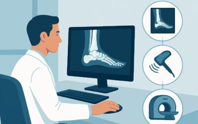
A 45-year-old patient, an avid tennis player, presents with six months of persistent, ulnar-sided wrist pain that worsens with rotation. They’ve tried rest, bracing, and NSAIDs with minimal relief. You ordered initial radiographs, which came back unremarkable, showing only minimal, nonspecific degenerative changes. The physical exam is suggestive of intra-articular pathology, but you need a definitive diagnosis to guide treatment. This is a common clinical crossroads where the next imaging choice is critical for identifying underlying ligamentous, cartilaginous, or osseous pathology missed by X-ray. For this specific scenario, the American College of Radiology (ACR) rates MR arthrography of the wrist as Usually Appropriate, providing the detailed anatomical map needed to solve the diagnostic puzzle.
Who Fits This Clinical Scenario?
This guidance is for adult patients with chronic wrist pain (typically lasting three months or longer) where initial imaging has been unrevealing. The key inclusion criteria are:
- Adult patient
- Chronic wrist pain (not acute trauma)
- Initial radiographs are normal or show only nonspecific findings, such as mild osteoarthritis, without evidence of a specific fracture, malunion, or advanced degenerative disease.
It is crucial to distinguish this presentation from similar but distinct clinical problems that follow different diagnostic pathways. This workflow does not apply if:
- The pain is acute following significant trauma. In that case, the primary concern is an occult fracture, and the imaging workup would be different.
- Radiographs show a known old scaphoid fracture. This presentation requires a dedicated workup to evaluate for nonunion, malunion, or osteonecrosis, which is a separate ACR variant.
- Symptoms are primarily neuropathic and localized to the median nerve distribution. If you suspect carpal tunnel syndrome, the appropriate next step is typically electrodiagnostic studies or dedicated nerve imaging, another distinct scenario.
- The primary suspicion is tendon injury or tenosynovitis. While MRI is still often used, the specific protocol and differential diagnosis differ from an intra-articular workup.
Correctly identifying your patient within this specific scenario ensures the next imaging study is optimized to answer the relevant clinical question.
What Diagnoses Are You Working Up in This Scenario?
When wrist radiographs are normal in the setting of chronic pain, the differential shifts from overt fractures and advanced arthritis to more subtle intra-articular pathologies. The advanced imaging study is intended to visualize the soft tissue and cartilaginous structures that X-rays cannot.
A primary consideration, especially with ulnar-sided pain, is a Triangular Fibrocartilage Complex (TFCC) tear. The TFCC is a key stabilizing structure of the distal radioulnar joint (DRUJ), and injuries are a frequent cause of chronic, activity-related wrist pain. These tears are often degenerative or post-traumatic and are invisible on plain films.
Another common cause of wrist pain and instability is an intrinsic ligament tear, most notably of the scapholunate (SL) or lunotriquetral (LT) ligaments. A complete tear of the SL ligament can lead to debilitating scapholunate advanced collapse (SLAC) wrist arthritis over time. Early detection with advanced imaging is critical.
Chondromalacia or early cartilage damage is also on the differential. Radiographs are insensitive to cartilage loss until joint space narrowing is significant. Advanced imaging can detect cartilage thinning, fibrillation, or full-thickness defects long before they are apparent on an X-ray, identifying an early source of mechanical pain.
Less common but highly consequential is avascular necrosis (AVN) of a carpal bone, particularly Kienböck disease (AVN of the lunate). In its early stages, the lunate may appear normal on radiographs, but MRI can detect the characteristic loss of blood supply and bone marrow edema, allowing for early intervention to prevent carpal collapse.
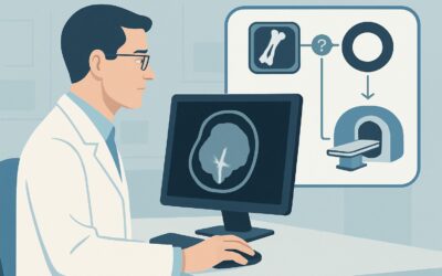
Why Is MR Arthrography the Recommended Study for This Presentation?
For an adult with chronic wrist pain and unrevealing radiographs, both MR arthrography wrist and MRI wrist without IV contrast are rated as Usually Appropriate by the ACR. However, MR arthrography is often favored for its superior ability to detect subtle tears of the intrinsic ligaments and TFCC.
The procedure involves injecting a dilute gadolinium-based contrast agent directly into the radiocarpal joint under fluoroscopic or ultrasound guidance. This distends the joint capsule and forces contrast into any communicating tears, making them highly conspicuous on the subsequent MRI images. This technique is particularly sensitive for identifying partial-thickness tears and perforations of the TFCC and intercarpal ligaments, which might be missed on a non-contrast study.
While a non-contrast MRI is also an excellent, non-invasive choice, its diagnostic accuracy for small ligamentous perforations may be lower. It remains highly effective for diagnosing other conditions on the differential, such as avascular necrosis, occult fractures, bone marrow edema, and ganglion cysts. The choice between the two often depends on the specific clinical suspicion and patient factors.
How do alternative studies compare?
- Ultrasound (US) wrist: Rated May be appropriate. Ultrasound is non-invasive and allows for dynamic assessment, which can be valuable for evaluating tendon subluxation or ligamentous instability under stress. However, it is highly operator-dependent, has a limited field of view, and provides a less comprehensive assessment of deep intra-articular structures like cartilage and bone marrow compared to MRI.
- CT arthrography wrist: Also rated May be appropriate. Like MR arthrography, it is excellent for detecting ligamentous and TFCC tears. Its primary drawbacks are the use of ionizing radiation (☢ <0.1 mSv) and inferior soft-tissue contrast resolution compared to MRI, making it less ideal for evaluating bone marrow pathology like AVN.
Ultimately, MR arthrography provides the most comprehensive evaluation for the most common differential diagnoses in this scenario, with no ionizing radiation (0 mSv).
Once you’ve decided on an MRI, our protocol guide covers the technical details, contrast considerations, and key reading principles. For a deeper dive, see: MRI Wrist/Hand.
What’s Next After MR Arthrography? Downstream Workflow
The results of the MR arthrogram will guide the subsequent management plan, branching into surgical, medical, or conservative pathways.
- If the study is positive for a significant TFCC or ligament tear (e.g., scapholunate, lunotriquetral): The next step is typically a referral to a hand surgeon or a sports medicine specialist. Depending on the tear’s severity, patient age, and activity level, treatment options range from conservative management with splinting and targeted physical therapy to surgical intervention, such as arthroscopic debridement or repair.
- If the study is positive for avascular necrosis (Kienböck disease): An urgent referral to a hand surgeon is warranted. Management is stage-dependent and aims to preserve wrist function and prevent carpal collapse. Options may include revascularization procedures, osteotomies, or, in advanced cases, carpal fusion.
- If the study is negative: A negative, high-quality MR arthrogram makes a significant intra-articular structural cause of pain much less likely. The focus should shift to extra-articular causes. This may prompt a re-evaluation for tenosynovitis, early inflammatory arthritis (requiring rheumatology consultation), or a neuropathic pain source. The clinical workup may now align more closely with the ACR scenario for suspected tendon injury.
- If the study is indeterminate or shows only nonspecific findings: Clinical correlation is paramount. The findings should be discussed with the reporting musculoskeletal radiologist. A diagnostic/therapeutic injection into a specific joint or tendon sheath can sometimes help isolate the pain generator and guide further treatment.
Pitfalls to Avoid (and When to Get Help)
Navigating the workup for chronic wrist pain requires careful consideration to avoid common diagnostic missteps.
1. Vague Requisitions: Ordering an “MRI wrist” with “chronic pain” as the only indication is a missed opportunity. Be specific. A history of “ulnar-sided wrist pain, worse with forearm rotation, rule out TFCC tear” allows the radiologist to tailor the protocol and focus their search.
2. Choosing Non-Arthrography for a Suspected Ligament Tear: While a non-contrast MRI is rated Usually Appropriate, if your pre-test suspicion for a small TFCC or intercarpal ligament perforation is high, MR arthrography offers the highest sensitivity and may prevent a false-negative result.
3. Ignoring Patient Contraindications: MR arthrography is an invasive procedure. Remember to screen for contrast allergies, renal insufficiency (for gadolinium), and active infection. It also may not be suitable for patients with severe anxiety or an inability to remain still.
4. Overlooking the Kinetic Chain: Wrist pain can be referred from the elbow, shoulder, or cervical spine. If a comprehensive wrist workup is negative, consider expanding the physical examination and differential to include more proximal pathology.
If the diagnosis remains elusive after advanced imaging or if findings suggest a systemic process like inflammatory arthritis, escalate by consulting with a musculoskeletal radiologist, hand surgeon, or rheumatologist.
Related ACR Topics and Tools
This article focuses on a single, common clinical scenario. For a comprehensive overview of imaging for all presentations of chronic hand and wrist pain, see our parent guide. It provides a hub-and-spoke model to help you navigate the full ACR Appropriateness Criteria topic.
For breadth across all scenarios in Chronic Hand and Wrist Pain, see our parent guide: Chronic Hand and Wrist Pain: ACR Appropriateness Decoded.
To explore other scenarios, optimize imaging techniques, or discuss radiation dose with patients, these GigHz resources can help:
Frequently Asked Questions
Is a non-contrast MRI a reasonable alternative to MR arthrography in this scenario?
Yes, an MRI of the wrist without IV contrast is also rated ‘Usually Appropriate’ by the ACR. It is an excellent non-invasive option that is highly effective for diagnosing conditions like avascular necrosis, bone marrow edema, and most soft tissue pathologies. However, MR arthrography may be slightly more sensitive for detecting small or partial-thickness tears of the TFCC and intrinsic ligaments.
Why not just order a CT scan instead of an MRI?
CT arthrography is rated ‘May be appropriate’ and is very good for evaluating ligaments, but it involves ionizing radiation and provides less detail of the bone marrow and soft tissues compared to MRI. A standard CT without contrast is generally not sufficient for evaluating the primary differential diagnoses in this scenario, which involve cartilage and ligaments.
What are the options if my patient is claustrophobic and cannot tolerate an MRI?
First, discuss options like open MRI (if available and of sufficient diagnostic quality) or the possibility of conscious sedation with the imaging facility. If an MRI is truly not feasible, CT arthrography becomes the next best alternative for a detailed intra-articular evaluation, though it comes with the trade-off of ionizing radiation.
How does a finding of ‘nonspecific arthritis’ on the initial radiograph affect the imaging choice?
It doesn’t change the recommendation. This clinical scenario explicitly includes patients whose radiographs show nonspecific arthritis. The purpose of the advanced imaging, like an MR arthrogram, is to determine if there is a specific, treatable cause for the patient’s pain (e.g., a ligament tear) beyond the mild, often asymptomatic degenerative changes seen on the X-ray.
When should I consider ordering an ultrasound first?
Ultrasound is rated ‘May be appropriate’ and can be a useful first-line advanced imaging test if your clinical suspicion is focused on a specific, superficial structure. For example, it is excellent for evaluating for tenosynovitis, a ganglion cyst, or performing a dynamic assessment of a tendon. For a comprehensive evaluation of intra-articular structures like the TFCC, cartilage, and bone marrow, MRI is superior.
Reviewed by Pouyan Golshani, MD, Interventional Radiologist — May 30, 2026