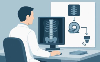
What Is the Best Imaging for Chronic Elbow Pain Refractory to Treatment?

A 48-year-old patient, an avid weekend tennis player, presents to your clinic with six months of persistent lateral elbow pain. They’ve diligently tried physical therapy, bracing, and activity modification with minimal relief. Your clinical suspicion is high for chronic lateral epicondylalgia, possibly with an underlying tendon tear, but their initial radiographs were unremarkable. You’re now faced with the decision of which advanced imaging study will best clarify the diagnosis and guide the next phase of treatment. This article provides a step-by-step workflow for this specific scenario, grounded in the American College of Radiology (ACR) Appropriateness Criteria. For this presentation, an Ultrasound (US) of the elbow is rated Usually Appropriate.
Who Fits This Clinical Scenario for Refractory Elbow Pain?
This guidance is tailored for a specific patient population. The recommendations apply when the clinical picture includes:
- Chronic Symptoms: The elbow pain has persisted for weeks to months.
- Refractory to Conservative Care: The patient has already undergone a trial of empirical treatments such as rest, nonsteroidal anti-inflammatory drugs (NSAIDs), physical therapy, or bracing without significant improvement.
- Primary Suspicion of Tendon Pathology: The history and physical exam point strongly toward lateral or medial epicondylalgia (also known as epicondylitis or tendinosis) or a tear of the common extensor or flexor tendons.
- Unremarkable Radiographs: Initial X-rays have already been performed and are either normal or show only nonspecific findings like minor calcifications, effectively ruling out significant osseous pathology.
It is critical to distinguish this scenario from similar presentations that follow different diagnostic pathways. This workflow is not intended for:
- Initial Imaging: Patients presenting with chronic elbow pain for the first time who have not yet had radiographs.
- Mechanical Symptoms: Patients with locking, clicking, or a true loss of range of motion, which suggests an intra-articular problem like a loose body or cartilage damage.
- Suspected Nerve Abnormality: Patients with primary neurologic symptoms like paresthesias or weakness in a specific nerve distribution (e.g., ulnar or radial nerve), which points toward a nerve entrapment syndrome.
What Diagnoses Are You Working Up in Refractory Epicondylalgia?
When a patient’s elbow pain persists despite conservative measures, imaging is used to confirm the diagnosis, assess the severity of the pathology, and rule out other causes. The differential diagnosis in this specific scenario is focused on the peritendinous soft tissues.
Lateral Epicondylitis (Tennis Elbow)
This is the most common cause of lateral elbow pain, involving tendinosis of the common extensor tendon origin at the lateral epicondyle. Imaging aims to identify tendon thickening, hypoechoic changes indicative of mucoid degeneration, and neovascularity on color Doppler. The goal is to confirm tendinosis and assess for any superimposed tearing.
Medial Epicondylitis (Golfer’s Elbow)
This is the medial counterpart to tennis elbow, affecting the common flexor tendon origin. The underlying pathology is the same—tendinosis and potential tearing—but located on the medial side of the elbow. The clinical exam typically localizes the pain, guiding the imaging evaluation.
Common Extensor or Flexor Tendon Tear
This represents a progression from simple tendinosis to structural failure of the tendon. Tears can be interstitial (within the substance of the tendon), partial-thickness (affecting the articular or bursal surface), or full-thickness. Advanced imaging is essential to determine the presence, size, and location of a tear, as this information directly influences whether treatment remains conservative or shifts toward interventions like percutaneous procedures or surgical repair.
Ligamentous Injury
Less commonly, chronic elbow pain can stem from an injury to the collateral ligaments, particularly the radial or ulnar collateral ligament complexes. These injuries can mimic the symptoms of epicondylalgia, and advanced imaging can be crucial in differentiating between the two.
Why Is Ultrasound the Recommended Next Step for Suspected Tendon Injury?
For a patient with refractory epicondylalgia and normal radiographs, the ACR designates both US elbow and MRI elbow without IV contrast as Usually Appropriate. While both are excellent choices, ultrasound often serves as the ideal first-line advanced imaging modality for several key reasons.
Ultrasound offers superb spatial resolution of superficial soft tissues, allowing for detailed evaluation of tendon architecture. It can readily identify tendon thickening, fiber disruption, calcifications, and fluid collections characteristic of tendinosis and tears. A major advantage of US is its ability to perform dynamic imaging. A skilled sonographer can have the patient move their elbow and wrist during the scan, assessing for tendon subluxation or impingement in real-time—a capability static imaging like MRI cannot replicate. Furthermore, US can be used to guide therapeutic injections in the same session, streamlining patient care.
MRI without contrast is an equally valid and powerful alternative. It is less operator-dependent than US and provides a more global view of the elbow, including bone marrow and deeper soft tissues that may be outside the ultrasound field of view. It is an excellent choice if the diagnosis is unclear, if there is suspicion of coexisting intra-articular pathology, or if a high-quality musculoskeletal ultrasound is not available.
Other imaging modalities are rated lower for this specific clinical question:
- MR Arthrography is rated Usually Not Appropriate. This invasive study involves injecting contrast directly into the joint and is designed to evaluate intra-articular structures like cartilage and the joint capsule, which are not the primary concern here.
- CT scans (with or without contrast) are also Usually Not Appropriate. CT provides exceptional bone detail but has poor soft-tissue contrast for evaluating tendons. It also exposes the patient to ionizing radiation (Relative Radiation Level ☢☢), making it unsuitable for a primary tendon workup.
What’s Next After an Elbow Ultrasound? Downstream Workflow
The results of the elbow ultrasound will guide the subsequent clinical management. The decision tree typically branches based on whether the findings confirm the suspected diagnosis, are negative, or reveal an unexpected pathology.
If the US is positive for epicondylitis or a low-grade tear:
This result confirms the clinical diagnosis and provides objective evidence of the tendon pathology. Management may continue with advanced conservative therapies, such as formal physical therapy focused on eccentric loading, or proceed to ultrasound-guided interventions. These can include corticosteroid injections for short-term relief or biologic injections (e.g., platelet-rich plasma) aimed at promoting tissue healing.
If the US is positive for a high-grade partial or full-thickness tear:
Identifying a significant structural tear is a critical finding that often changes the treatment plan. This result typically prompts a referral to an orthopedic surgeon to discuss options for surgical repair, especially for active individuals or those who have failed all non-operative treatments.
If the US is negative or indeterminate:
When a high-quality ultrasound is normal but the patient’s symptoms and clinical signs strongly suggest pathology, the next logical step is to consider the other Usually Appropriate study: an MRI of the elbow without IV contrast. MRI may detect subtle interstitial tears, bone marrow edema, or other soft-tissue abnormalities not visualized on US. If both US and MRI are negative, the focus should shift back to non-imaging-based diagnoses, such as referred pain or central sensitization.
Common Pitfalls in Imaging Refractory Elbow Pain
Navigating the workup for chronic elbow pain requires avoiding several common diagnostic traps. First, do not anchor on a “normal” radiograph as evidence against significant pathology; X-rays are insensitive to the tendinosis and tears that cause this clinical picture. Second, recognize the operator-dependent nature of ultrasound. If the results of a US exam do not align with a strong clinical suspicion, consider obtaining a second opinion from an experienced musculoskeletal sonographer or proceeding to MRI.
Another pitfall is ordering the wrong advanced study. For suspected tendon pathology, CT provides limited useful information and unnecessary radiation. Similarly, ordering an MR arthrogram for suspected epicondylitis is an invasive procedure that does not target the primary area of concern. Adhering to the ACR criteria helps ensure the right test is chosen for the right clinical question. If red flag symptoms such as fever, overlying erythema, or rapidly progressive neurologic deficits develop, escalate care immediately for a workup of infection or an acute compressive neuropathy.
Related ACR Topics and Tools
This article focuses on a single, common clinical scenario. For a comprehensive overview of all variants and imaging modalities related to chronic elbow pain, or to explore adjacent clinical questions, the following resources are available.
- For breadth across all scenarios in Chronic Elbow Pain, see our parent guide: Chronic Elbow Pain: ACR Appropriateness Decoded.
- To explore other clinical presentations, use the ACR Appropriateness Criteria Lookup.
- For details on imaging techniques, consult the Imaging Protocol Library.
- To discuss radiation exposure with patients, use the Radiation Dose Calculator.
Frequently Asked Questions
Why is MRI without contrast also ‘Usually Appropriate’ if ultrasound is so effective?
MRI without contrast is also rated ‘Usually Appropriate’ because it provides excellent, non-operator-dependent anatomical detail of tendons, ligaments, bone, and muscle. It’s a superb alternative if high-quality musculoskeletal ultrasound is unavailable, if the ultrasound results are inconclusive, or if there’s a broader differential diagnosis that includes potential bone marrow or deep soft-tissue pathology that US might miss.
Should I order imaging with contrast for suspected epicondylitis?
No, for this specific scenario, both MRI with and without IV contrast and CT with IV contrast are rated ‘Usually Not Appropriate’. The primary pathology in epicondylitis (tendinosis and tearing) is well-visualized on non-contrast sequences. IV contrast does not typically add diagnostic value for this indication and introduces unnecessary cost and potential risks (e.g., gadolinium exposure or contrast reaction).
What if the patient’s pain is diffuse and not clearly localized to the medial or lateral epicondyle?
If the pain is diffuse, the pre-test probability of epicondylitis decreases, and the differential diagnosis broadens. In this case, MRI without contrast may be a better initial choice than ultrasound. MRI provides a more comprehensive overview of the entire elbow joint, including intra-articular structures, bone marrow, and surrounding musculature, making it better suited for a less-localized clinical presentation.
Can ultrasound differentiate between tendinosis and a partial-thickness tear?
Yes, a high-quality musculoskeletal ultrasound can reliably differentiate between the two. Tendinosis typically appears as tendon thickening and hypoechogenicity (darker appearance) without a discrete fiber defect. A partial-thickness tear will show a focal, anechoic or hypoechoic cleft or defect within the tendon fibers, indicating structural disruption.
If the initial radiographs showed some calcification near the epicondyle, does that change the next step?
No, the presence of calcific tendinopathy on radiographs does not change the recommended next step. This finding actually increases the suspicion for chronic tendinosis. An ultrasound would still be the ‘Usually Appropriate’ next study to assess the integrity of the rest of the tendon, look for associated tears, and potentially guide a barbotage procedure to break up the calcium deposit if indicated.
Reviewed by Pouyan Golshani, MD, Interventional Radiologist — May 30, 2026