What Is the Best Imaging for Follow-Up of Liver-Dominant Pancreatic Neuroendocrine Tumors?

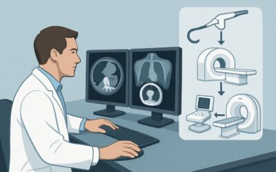
It’s Tuesday afternoon, and you’re reviewing the follow-up plan for a 58-year-old patient with a well-differentiated pancreatic neuroendocrine tumor (pNET) who recently completed a cycle of liver-directed therapy. The bulk of their disease is in the liver, and the key clinical question is whether the treatment was effective and if any new lesions have appeared. Choosing the right imaging modality is critical for guiding the next steps in management, balancing diagnostic accuracy with the need for repeated scans over the patient’s lifetime. This article details the clinical workflow for this specific scenario: follow-up imaging for an adult with treated, liver-dominant pNET. Based on the American College of Radiology (ACR) Appropriateness Criteria, MRI abdomen and pelvis without and with IV contrast is rated Usually Appropriate for this indication.
Who Fits This Clinical Scenario?
This guidance applies to adult patients with a known, pathologically confirmed pancreatic neuroendocrine tumor where the predominant disease burden is metastatic to the liver. Critically, this workflow is for follow-up after treatment, such as trans-arterial chemoembolization (TACE), radioembolization, ablation, or systemic therapy. The primary goal of imaging is to assess treatment response, detect recurrence or progression within the liver, and identify any new extrahepatic disease.
This article does not apply to several similar-sounding but distinct clinical situations, which have their own recommended imaging pathways:
- Initial Staging: Patients newly diagnosed with a pNET who have not yet undergone treatment require a comprehensive staging evaluation to determine the full extent of disease.
- Post-Surgical Surveillance: Patients who have undergone complete surgical resection of their primary tumor and any known metastases, and who have no suspected or known recurrence, follow a different surveillance protocol.
- Non-Liver Dominant Disease: Patients whose primary disease burden is extrahepatic (e.g., extensive nodal, bone, or peritoneal disease) may require a different imaging strategy.
- Follow-up of Untreated Disease: Patients with known disease who are on a “watch and wait” or surveillance protocol without active treatment have different imaging considerations.
Correctly identifying your patient’s scenario ensures the most appropriate and highest-value imaging is selected.
What Diagnoses Are You Working Up in This Scenario?
In the context of post-treatment follow-up for liver-dominant pNETs, the imaging “workup” is less about a new diagnosis and more about characterizing the status of known disease. The key questions you are trying to answer with the imaging study drive the choice of modality.
Treatment Response: The most immediate goal is to determine if the treated liver lesions have responded. This isn’t just about size. Imaging can reveal necrosis, decreased vascularity, or other signs of successful therapy, which may precede a change in lesion diameter. Distinguishing viable, enhancing tumor from post-treatment scar or necrosis is a primary objective.
Disease Progression: Conversely, you are looking for evidence of progression. This can manifest as an increase in the size of treated lesions, the development of new enhancement within a previously treated lesion, or, most commonly, the appearance of new hepatic or extrahepatic metastases. Identifying progression early is crucial for deciding whether to change or escalate therapy.
Stable Disease: Many patients will demonstrate stable disease, where there is no significant change in tumor burden. This finding is also clinically actionable, as it typically supports continuing the current management plan and scheduling the next surveillance scan.
Treatment-Related Complications: Liver-directed therapies can have complications. Imaging can help identify potential issues such as abscess formation, biliary obstruction, or non-target embolization. While less common, these are consequential findings that can alter patient management significantly.
Why Is MRI Abdomen and Pelvis Without and With IV Contrast the Recommended Study?
For assessing treated liver-dominant pNETs, MRI provides superior diagnostic information compared to other modalities, earning its Usually Appropriate rating from the ACR. The rationale is multifaceted, focusing on soft-tissue characterization, sensitivity for new lesions, and radiation safety.
The primary advantage of MRI is its exceptional soft-tissue contrast resolution. This allows for detailed evaluation of liver parenchyma and metastatic deposits, making it highly effective at distinguishing viable, enhancing tumor from post-treatment changes like necrosis or fibrosis. Specific sequences, like diffusion-weighted imaging (DWI), can provide functional information about tumor cellularity, often showing treatment response before size changes are apparent on standard sequences.
When comparing MRI to alternatives, the trade-offs become clear:
- CT abdomen and pelvis without and with IV contrast is also rated Usually Appropriate. It is an excellent modality, particularly for assessing the hypervascular nature of pNET metastases in the arterial phase. However, it involves significant ionizing radiation (☢☢☢☢ 10-30 mSv). Given that these patients require serial imaging for years, the cumulative radiation dose is a major consideration. MRI (O 0 mSv) avoids this risk entirely, making it the preferred choice for routine follow-up.
- DOTATATE PET/CT is rated May be appropriate. This functional imaging study is invaluable for assessing somatostatin receptor (SSTR) expression across the entire body and is critical for selecting patients for peptide receptor radionuclide therapy (PRRT). However, for routine morphological assessment of treatment response in the liver, its spatial resolution is lower than MRI or CT. It is often used adjunctively if there is a discrepancy on anatomic imaging or to re-evaluate SSTR status before a change in therapy, not as the primary tool for every follow-up scan.
For these reasons, MRI is the cornerstone of follow-up. When ordering, it is crucial to request a multiphasic liver protocol that includes pre-contrast, late arterial, portal venous, and delayed phases, as well as DWI sequences, to maximize the diagnostic yield.
What’s Next After MRI? Downstream Workflow
The results of the follow-up MRI will directly guide the subsequent clinical management. The decision tree branches based on whether the findings show response, stability, or progression.
If the MRI shows a good treatment response or stable disease: This is an encouraging result. The typical next step is to continue with the current management plan and schedule the next interval follow-up scan. The frequency of imaging depends on the tumor grade, disease burden, and specific therapy, but often ranges from every 3 to 12 months.
If the MRI shows unequivocal disease progression: This finding necessitates a re-evaluation of the treatment strategy. This could involve a multidisciplinary tumor board discussion to consider options such as switching to a different systemic agent, initiating a new line of liver-directed therapy for new or growing lesions, or evaluating for PRRT if the patient is a candidate and has not received it previously.
If the MRI is indeterminate or equivocal: Sometimes a finding is not clearly benign or malignant, such as a new, tiny liver lesion or an atypical change in a treated lesion. In this situation, the downstream workflow may involve a short-interval follow-up MRI (e.g., in 2-3 months) to assess for change. Alternatively, if a change in management hinges on the finding, a problem-solving study like a DOTATATE PET/CT can be used to determine if the lesion is SSTR-avid, suggesting active neuroendocrine tumor. In rare cases, a biopsy may be required.
Pitfalls to Avoid (and When to Get Help)
Several common pitfalls can compromise the value of follow-up imaging in this scenario. Awareness of these issues can help ensure optimal patient care.
- Inconsistent Modality: Alternating between CT and MRI for follow-up makes direct comparison of lesion size and characteristics difficult. Sticking with one modality, preferably MRI, provides the most reliable assessment of change over time.
- Using a Generic Protocol: Ordering a “routine” abdomen MRI without specifying a multiphasic liver protocol may result in a non-diagnostic study. Always ensure the protocol is optimized for detecting and characterizing hypervascular liver metastases.
- Over-reliance on RECIST: Relying solely on size-based criteria (Response Evaluation Criteria in Solid Tumors) can be misleading. A treated lesion may not shrink but can become necrotic and non-enhancing, representing an excellent response. The radiology report should incorporate changes in morphology and enhancement, not just size.
- Forgetting Extrahepatic Sites: While the disease is liver-dominant, progression can occur elsewhere. Ensure the ordered study (e.g., MRI of the abdomen and pelvis) and the radiologist’s review covers common sites of extrahepatic disease.
If imaging findings are complex or discordant with the clinical picture, discussion at a multidisciplinary neuroendocrine tumor board is the best path to an integrated and expert-guided management plan.
Related ACR Topics and Tools
For a comprehensive overview of imaging across all clinical presentations of this condition, please consult the parent topic article. Additional GigHz tools can help you apply these standards in your daily practice.
- For breadth across all scenarios in Staging and Follow-up of Pancreatic Neuroendocrine Tumors, see our parent guide: Staging and Follow-up of Pancreatic Neuroendocrine Tumors: ACR Appropriateness Decoded.
- To explore other clinical scenarios, use the ACR Appropriateness Criteria Lookup.
- For technical details on performing the recommended study, see the Imaging Protocol Library.
- To discuss cumulative radiation exposure with patients when considering CT, use the Radiation Dose Calculator.
Frequently Asked Questions
Why is MRI preferred over CT for follow-up if both are rated ‘Usually Appropriate’?
The primary reason is radiation safety. Patients with pNETs often require many scans over their lifetime. MRI uses no ionizing radiation, making it the safer choice for serial follow-up. Additionally, MRI offers superior soft-tissue contrast and functional information (like DWI) that can be more sensitive for detecting treatment response and small new lesions compared to CT.
When should I order a DOTATATE PET/CT instead of an MRI for follow-up?
A DOTATATE PET/CT is not the primary tool for routine morphological follow-up of liver lesions. It should be considered when there is a need to assess whole-body somatostatin receptor (SSTR) status, such as before initiating PRRT, or as a problem-solving tool when MRI findings are equivocal and knowing a lesion’s SSTR-avidity would change management.
What if my patient has a contraindication to MRI, like an incompatible pacemaker?
In cases where MRI is contraindicated, a multiphasic CT of the abdomen and pelvis without and with IV contrast is the best alternative. It is also rated ‘Usually Appropriate’ by the ACR and provides excellent anatomic detail, especially for hypervascular liver metastases, though it involves radiation exposure.
How often should follow-up imaging be performed?
The optimal frequency of follow-up imaging is not standardized and depends on multiple factors, including the tumor grade (G1, G2, G3), the extent of disease, the specific treatment received, and the patient’s clinical status. Guidelines from organizations like NCCN typically suggest intervals ranging from every 3 to 12 months, a decision best made in consultation with the patient’s oncology team.
Does this guidance apply to poorly differentiated neuroendocrine carcinomas (NECs)?
This guidance is primarily for well-differentiated neuroendocrine tumors (NETs). Poorly differentiated, high-grade neuroendocrine carcinomas (NECs) are biologically different, often more aggressive, and typically do not express somatostatin receptors. They are often followed with FDG-PET/CT, as they are more metabolically active. This specific ACR variant focuses on the more common well-differentiated pNETs.
Reviewed by Pouyan Golshani, MD, Interventional Radiologist — May 30, 2026