What Is the Best Imaging for Local Recurrence of a Soft Tissue Tumor?

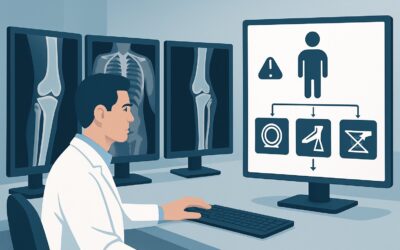
A 58-year-old patient is in your clinic for a routine 2-year follow-up after a wide local excision of a myxofibrosarcoma from his posterior thigh. He is asymptomatic, and the surgical site is well-healed on physical exam. Per the surveillance protocol, it is time for imaging to assess for local recurrence. You need to decide on the most effective study to detect a small, non-palpable recurrence within the complex post-surgical and post-radiation tissue bed. This article details the clinical workflow for this specific scenario, guiding you to the most appropriate imaging choice. According to the American College of Radiology (ACR) Appropriateness Criteria, MRI of the area of interest without and with IV contrast is rated Usually Appropriate for this indication.
Who Fits This Clinical Scenario for Soft Tissue Tumor Surveillance?
This guidance applies specifically to patients undergoing planned surveillance for local recurrence of a previously treated malignant or aggressive primary soft tissue tumor. The key inclusion criteria are a confirmed history of a soft tissue malignancy (e.g., sarcoma) that has been treated with curative intent (typically surgery with or without radiation) and a current clinical status of being asymptomatic at the primary tumor site.
It is critical to distinguish this scenario from several related but distinct clinical questions that require different imaging pathways:
- Initial Staging: This workflow is not for the initial workup of a newly diagnosed tumor. Staging for pulmonary or extrapulmonary metastasis follows a different set of recommendations.
- Primary Bone Tumors: Surveillance for a primary bone tumor, such as an osteosarcoma or Ewing sarcoma, involves different considerations for local recurrence and is covered under a separate ACR variant.
- Symptomatic Patients: If the patient presents with a new palpable lump, pain, or other symptoms at the surgical site, the workup shifts from routine surveillance to a diagnostic evaluation, which may alter the urgency and choice of imaging.
- Distant Metastasis Surveillance: This article focuses solely on local recurrence at the primary site. The strategy for monitoring for distant disease (e.g., in the lungs) is a separate clinical question.
What Diagnoses Are You Working Up in This Scenario?
While the primary goal of surveillance is to detect a single entity—recurrent cancer—the imaging must reliably differentiate it from the expected and benign changes that occur after treatment. The differential in this context is less about multiple possible diseases and more about distinguishing recurrence from its mimics.
Local Tumor Recurrence This is the principal target of surveillance. A local recurrence is the regrowth of the tumor at or immediately adjacent to the original tumor site. It can manifest as a discrete nodule or as a more subtle, infiltrative process. Early detection is paramount, as it offers the best chance for successful salvage therapy, which may include re-excision, further radiation, or systemic treatment.
Post-Surgical and Post-Radiation Changes This is the most common confounder and the primary reason high-quality imaging is essential. The surgical bed will contain scar tissue (fibrosis), which can be nodular and mimic a tumor. Other common findings include seromas, hematomas, fat necrosis, and muscle atrophy. Radiation therapy adds another layer of complexity, causing edema, inflammation, and eventual fibrosis that can obscure or be mistaken for recurrence.
Benign Post-Treatment Masses Less commonly, a new mass in the surgical bed may be benign. Suture granulomas, for instance, are inflammatory reactions to suture material that can form a discrete, enhancing nodule. An organized hematoma or a myositis ossificans-like process can also develop. While less frequent, these must be distinguished from a true recurrence to avoid unnecessary invasive procedures.
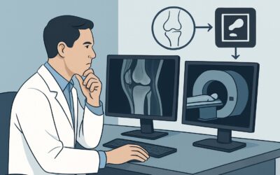
Why Is MRI Without and With Contrast the Recommended Study for Local Recurrence?
The ACR designates MRI of the area of interest without and with IV contrast as Usually Appropriate because of its unparalleled ability to characterize soft tissues and differentiate between malignant and benign post-treatment changes.
The core strength of MRI is its superior soft-tissue contrast resolution. It can distinguish subtle differences between muscle, fat, scar tissue, edema, and recurrent tumor far better than any other modality. T1-weighted sequences are excellent for anatomical detail and identifying fat, while fluid-sensitive sequences like T2-weighted or STIR (Short Tau Inversion Recovery) imaging are highly effective at detecting edema and abnormal fluid collections, which can be early signs of recurrence.
The administration of a gadolinium-based intravenous contrast agent is critical. Recurrent tumors are typically hypervascular and demonstrate avid enhancement, often earlier and more intensely than surrounding scar tissue. Fibrotic scar tissue tends to enhance more slowly and less robustly. Comparing pre-contrast images to dynamic post-contrast sequences allows the radiologist to assess the vascularity and enhancement pattern of any suspicious area, which is often the key to making the correct diagnosis. A baseline MRI performed 3-6 months after treatment completion is invaluable, as it establishes the expected post-treatment appearance against which all future surveillance scans can be compared.
Why are other studies rated lower for this specific scenario?
- CT with IV contrast is rated May be appropriate. While it can detect larger recurrences, its soft tissue resolution is significantly inferior to MRI. Differentiating a small recurrent nodule from post-surgical scar is extremely challenging with CT, leading to lower sensitivity and specificity. It remains a viable alternative if MRI is absolutely contraindicated (e.g., incompatible metallic implants, severe claustrophobia). Note that this study involves ionizing radiation (dose varies).
- Ultrasound (US) of the area of interest is rated May be appropriate (Disagreement). The “Disagreement” reflects the modality’s significant limitations in this role. While US is excellent for superficial, palpable lesions and can guide biopsies, it is highly operator-dependent, has a limited field of view, and struggles to penetrate deep tissues or areas with significant scarring. It is generally not considered a primary tool for comprehensive surveillance of a deep surgical bed.
The ACR also rates MRI of the area of interest without IV contrast as Usually Appropriate. This is an important option for patients with contraindications to gadolinium, such as severe renal impairment or a history of severe allergic reaction. While the lack of contrast reduces diagnostic confidence in differentiating scar from tumor, the morphological information from non-contrast sequences is still highly valuable and superior to other non-contrast modalities.
What Is the Downstream Workflow After a Surveillance MRI?
The results of the surveillance MRI will direct the next steps in patient management, typically falling into one of three pathways.
Positive for Suspected Recurrence If the MRI identifies a new or growing enhancing mass, or other features highly suspicious for local recurrence, the standard next step is a biopsy. An image-guided core needle biopsy (often using ultrasound or CT for guidance) is performed to obtain tissue for histopathologic confirmation. A confirmed recurrence triggers a multidisciplinary tumor board discussion involving surgical oncology, radiation oncology, and medical oncology to determine the optimal plan for salvage therapy.
Negative or Stable Post-Treatment Changes When the MRI shows no evidence of recurrence and demonstrates only expected post-treatment changes that are stable compared to prior scans, the patient can safely continue their established surveillance schedule. This typically involves a follow-up clinical visit and repeat imaging at a prescribed interval (e.g., 6 to 12 months), with the frequency depending on the original tumor’s grade, stage, and the time elapsed since treatment.
Indeterminate Finding Occasionally, the MRI may reveal an equivocal finding—an area of subtle new enhancement or a non-specific change that is not definitively benign or malignant. In this situation, management options include a short-interval follow-up MRI (e.g., in 3 months) to assess for stability or progression, which can often clarify the nature of the finding. Alternatively, if clinical suspicion is higher or waiting is undesirable, a functional imaging study like FDG-PET/CT (rated May be appropriate) can be used as a problem-solving tool. Metabolically active tumor recurrence will typically show avid FDG uptake, whereas benign scar tissue will not. If suspicion remains high despite these measures, a biopsy may still be warranted.
Common Pitfalls to Avoid in Soft Tissue Tumor Surveillance
Navigating surveillance requires careful attention to detail to maximize the benefit of imaging while avoiding errors.
- Forgetting the Baseline Scan: The single most important factor for accurate interpretation is comparison to a baseline post-treatment MRI, typically obtained 3-6 months after therapy completion. Without it, distinguishing early recurrence from expected scar is exceptionally difficult.
- Inconsistent Imaging Technique: Using different MRI protocols, scanners, or imaging centers for serial surveillance scans can introduce variability that makes subtle changes difficult to detect. Consistency is key.
- Over-reliance on a Single Sequence: A comprehensive MRI protocol is essential. A diagnosis should not be made based on a single finding, such as enhancement alone. The morphology on T1 and T2 images, signal characteristics, and comparison to prior studies are all crucial.
- Ignoring Clinical Context: The imaging findings must always be correlated with the patient’s history, the original tumor’s histology and grade, and the physical exam. A low-grade tumor is less likely to recur than a high-grade one, which influences the pre-test probability of any ambiguous finding.
If an indeterminate finding persists across multiple imaging studies or if there is a discrepancy between imaging and clinical findings, escalation to a multidisciplinary tumor board or consultation with a musculoskeletal radiologist specializing in sarcoma imaging is the appropriate next step.
Related ACR Topics and Tools
For a comprehensive overview of imaging for all related clinical presentations, please refer to the parent topic guide. Additional tools can help you apply these criteria and understand the technical aspects of the recommended studies.
- For breadth across all scenarios in Malignant or Aggressive Primary Musculoskeletal Tumor-Staging And Surveillance, see our parent guide: Malignant or Aggressive Primary Musculoskeletal Tumor-Staging And Surveillance: ACR Appropriateness Decoded.
- ACR Appropriateness Criteria Lookup: For searching adjacent or alternative clinical scenarios.
- Imaging Protocol Library: For detailed technical guidance on performing the recommended imaging studies.
- Radiation Dose Calculator: For discussing cumulative radiation exposure with patients when considering CT or PET/CT.
Frequently Asked Questions
How often should surveillance MRI be performed for a soft tissue sarcoma?
The frequency of surveillance imaging depends on the tumor’s grade, stage, and the time since initial treatment. Guidelines from organizations like the National Comprehensive Cancer Network (NCCN) typically recommend more frequent imaging (e.g., every 3-6 months) for the first 2-3 years, then decreasing frequency annually up to 5 or 10 years post-treatment. This should be tailored to the individual patient’s risk profile.
Is an MRI without contrast sufficient for surveillance if a patient has a gadolinium allergy?
Yes, the ACR rates MRI without IV contrast as ‘Usually Appropriate’ for this scenario. While contrast enhancement is very helpful for differentiating recurrence from scar, a high-quality non-contrast MRI with multiple sequences (T1, T2, STIR) still provides excellent soft tissue detail and is far superior to CT. It is a very strong alternative when gadolinium is contraindicated.
When should I consider a PET/CT instead of an MRI for local surveillance?
According to the ACR, FDG-PET/CT is rated ‘May be appropriate’. It is not a first-line surveillance tool for local recurrence but serves as an excellent problem-solving modality. Its primary role is in evaluating an indeterminate finding on MRI. The metabolic activity shown on PET can help differentiate active tumor from metabolically inert scar tissue. It is also used for systemic staging, not just local surveillance.
What if the patient has surgical hardware from the initial resection?
The presence of metallic hardware can create significant artifacts on MRI. Modern MRI techniques, such as metal artifact reduction sequences (MARS), can substantially mitigate these artifacts. It is crucial to communicate the presence and type of hardware to the radiology department when ordering the scan so they can optimize the protocol. If the artifact is too severe to allow for a diagnostic study, CT with IV contrast may be the best alternative.
Does this guidance apply to desmoid tumors (aggressive fibromatosis)?
Yes, although desmoid tumors do not metastasize, they are locally aggressive and have a high rate of local recurrence. Therefore, the surveillance strategy is similar to that for low-grade soft tissue sarcomas. MRI without and with contrast is the imaging modality of choice for monitoring the primary site after treatment.
Reviewed by Pouyan Golshani, MD, Interventional Radiologist — May 30, 2026