What Imaging Should You Order for Extremity Trauma in a Stable Polytrauma Patient?
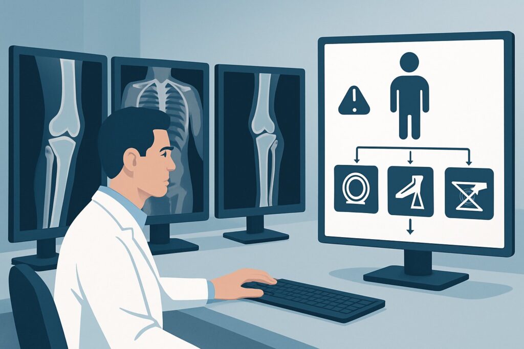
A 34-year-old arrives in the trauma bay after a high-speed motor vehicle collision. They are awake, alert, and hemodynamically stable, but have multiple abrasions and complain of severe right leg and left arm pain. As the primary clinician, you’ve completed your initial survey and stabilized the patient. Now you face a critical decision: what is the most appropriate initial imaging to evaluate their suspected extremity injuries without delaying the assessment for other, more occult thoracoabdominal trauma? This article provides a focused workflow for this exact scenario, walking through the differential, study rationale, and downstream decisions. According to the American College of Radiology (ACR) Appropriateness Criteria, for a stable adult with major blunt trauma and suspected extremity injury, a Radiography trauma series is Usually Appropriate.
Who Fits This Clinical Scenario?
This guidance applies specifically to an adult patient who has sustained major blunt trauma and is hemodynamically stable, with clinical suspicion for an extremity injury. “Major blunt trauma” typically implies a significant mechanism of injury, such as a motor vehicle crash, a fall from height, or a pedestrian being struck by a vehicle. “Hemodynamically stable” is a critical qualifier, meaning the patient has a normal blood pressure, heart rate, and is not in shock, allowing for a more deliberate imaging workup.
This workflow is NOT for:
- Hemodynamically unstable patients: These patients require a different, often more expedited, imaging protocol (like a FAST exam at the bedside followed by a rapid “pan-scan” CT) as their immediate life-threat is exsanguination. Their evaluation falls under a separate ACR variant.
- Patients with isolated extremity trauma: A person who, for example, twists their ankle playing sports without any other trauma does not fit the “major blunt trauma” context. Their workup is more focused.
- Patients whose primary suspected injury is elsewhere: If the leading concern is for facial, chest, or abdominal injury despite the presence of extremity pain, the imaging strategy should be guided by the ACR criteria for those specific scenarios, as the priorities for diagnosis change.
What Diagnoses Are You Working Up in This Scenario?
In a stable polytrauma patient with extremity pain, the imaging strategy is designed to rapidly identify or exclude several key injuries. The differential diagnosis guides the choice of the initial study.
Fractures: This is the most common and primary consideration. The goal is to identify fractures of the long bones (femur, tibia/fibula, humerus), pelvis, or smaller bones of the hands and feet. In the context of major trauma, identifying all fractures is crucial for stabilization and preventing further injury, such as neurovascular damage from unstable bone fragments.
Joint Dislocations: High-energy trauma can cause major joint dislocations, particularly of the hip, knee, or shoulder. These are orthopedic emergencies that require prompt reduction to restore blood flow and preserve joint function. Radiographs are highly effective at identifying dislocations.
Occult Injuries: A significant pitfall in polytrauma is the “distracting injury,” where a painful, obvious injury (like a femur fracture) masks the pain from a more subtle one (like a wrist or ankle fracture). A systematic radiographic survey helps mitigate this risk by imaging areas beyond the most obvious source of pain.
Associated Vascular Injury: While less common than fractures, vascular injuries are limb-threatening. Certain fracture patterns, like a knee dislocation or a displaced supracondylar femur fracture, have a high association with arterial injury (e.g., popliteal artery). While initial radiographs diagnose the bony injury, their findings often trigger a subsequent, more specific vascular imaging study.
Why Is a Radiography Trauma Series the Recommended Initial Study?
The ACR rates a Radiography trauma series as Usually Appropriate because it provides the best balance of diagnostic speed, availability, and utility for the most common and urgent diagnoses in this scenario. This series typically includes an AP pelvis, chest, and lateral cervical spine radiograph, with additional views of any symptomatic extremities.
Radiographs are excellent for the primary task: identifying fractures and dislocations. They are fast, can often be performed with portable machines in the trauma bay, and expose the patient to a relatively moderate amount of radiation (ACR Relative Radiation Level ☢☢☢). This allows for rapid orthopedic consultation and initial stabilization (splinting) while other aspects of the trauma workup proceed.
Two other studies are also rated Usually Appropriate: Radiography of the specific extremity area of interest and CT whole body with IV contrast. A focused extremity radiograph is essentially part of the trauma series. The whole-body CT (often called a “pan-scan”) is frequently performed in major trauma to evaluate for solid organ, great vessel, and spine injuries. While it can detect many extremity fractures, it is less sensitive for subtle, non-displaced, or articular-surface fractures compared to dedicated radiographs. Often, both are performed concurrently—the pan-scan for internal injuries and radiographs for a detailed extremity skeletal survey.
Why are other studies rated lower for the initial workup?
- CT of an extremity without IV contrast is rated Usually not appropriate. If the clinical question can be answered by non-contrast imaging, a radiograph is faster, cheaper, and uses less radiation. If cross-sectional imaging is truly needed for bony detail, it is often performed after initial radiographs to plan for surgery.
- US duplex Doppler of an extremity is rated Usually not appropriate as the first imaging test for a generalized suspicion of trauma. While it is the gold standard for evaluating a specific vascular injury, it should be ordered based on clinical signs (hard signs of vascular injury) or after a radiograph reveals a high-risk fracture pattern, not as a screening tool for all extremity pain.
What’s Next After Radiographs? Downstream Workflow
The results of the initial radiographs dictate the next steps in the patient’s care, creating a clear decision tree for the clinical team.
If radiographs are POSITIVE for fracture or dislocation: The immediate next step is consultation with orthopedic surgery. The patient will require stabilization, such as splinting or traction, and a plan for definitive management (e.g., closed reduction, operative fixation). For complex fractures, especially those involving a joint surface, the orthopedic surgeon may request a follow-up CT of the specific extremity to better characterize the fracture pattern and plan for surgery.
If radiographs are NEGATIVE but clinical suspicion remains high: A negative radiograph does not rule out all significant injury. If the patient has persistent, severe pain, deformity, or signs of neurovascular compromise, further investigation is mandatory. This is the point where a study rated May be appropriate, such as a CTA extremity area of interest with IV contrast, becomes critical, especially if there are hard or soft signs of a vascular injury. Alternatively, a non-contrast CT of the extremity may be ordered to look for an occult fracture not visible on plain films.
If radiographs reveal a high-risk injury pattern: Certain findings, such as a knee dislocation (even if spontaneously reduced) or a severely displaced fracture near a major artery, should immediately trigger a vascular assessment. This typically involves a physical exam for “hard signs” (e.g., absent pulses, expanding hematoma) and often proceeds directly to a CTA to rule out an arterial injury, which is a limb-threatening emergency.
Pitfalls to Avoid (and When to Get Help)
Navigating this scenario requires vigilance to avoid common diagnostic traps. Here are key pitfalls to watch for:
- Stopping the search after the first injury: In polytrauma, assume there is a second injury until proven otherwise. A painful femur fracture can easily distract both the patient and the clinician from a subtle wrist fracture. A systematic survey is essential.
- Ignoring the physical exam after imaging: A normal radiograph in the setting of a tense, swollen, and exquisitely painful limb should raise immediate concern for compartment syndrome, a clinical diagnosis that requires urgent surgical consultation (fasciotomy) and cannot be ruled out by imaging.
- Underestimating knee dislocations: Up to 50% of knee dislocations spontaneously reduce before the patient arrives at the hospital. A “normal” knee radiograph in a patient with a swollen, unstable knee and a high-energy mechanism requires a high index of suspicion for an occult dislocation and associated popliteal artery injury.
- Delaying vascular assessment: If hard signs of vascular injury are present (pulselessness, pallor, paresthesias, paralysis, poikilothermia, expanding hematoma), do not wait for advanced imaging to consult vascular surgery. This is a clinical decision that requires immediate escalation.
Related ACR Topics and Tools
This article covers one specific variant within the broader topic of Major Blunt Trauma. For a comprehensive overview of all related scenarios and to ensure you are applying the correct guidance, please consult the resources below.
- For breadth across all scenarios in Major Blunt Trauma, see our parent guide: Major Blunt Trauma: ACR Appropriateness Decoded.
- To explore other clinical presentations, use the ACR Appropriateness Criteria Lookup.
- For details on imaging techniques, visit the Imaging Protocol Library.
- To discuss radiation exposure with patients, the Radiation Dose Calculator can help frame the conversation.
Frequently Asked Questions
Why not just get a whole-body CT (‘pan-scan’) on every major trauma patient to cover the extremities too?
A whole-body CT is often performed in major trauma to assess for life-threatening injuries to the chest, abdomen, and pelvis. While it can identify many extremity fractures, it is less sensitive than dedicated radiographs for subtle, non-displaced, or complex joint fractures. Radiographs provide higher-resolution bony detail. The two studies are complementary and are often performed simultaneously in the initial workup.
If the patient is stable, can I wait to get the extremity radiographs?
While the patient is stable, delaying the diagnosis of fractures or dislocations can lead to complications like increased pain, swelling, and potential neurovascular compromise. Initial radiographs allow for prompt splinting and stabilization, which is a key part of early trauma care. They should be obtained as soon as practical after the primary and secondary surveys are complete.
When should I order a CTA of the extremity instead of a plain radiograph first?
A CTA should be considered as a primary or immediate follow-up study if there are ‘hard signs’ of vascular injury, such as absent distal pulses, an expanding hematoma, or a palpable thrill. In these cases, the concern for a limb-threatening vascular injury outweighs the need to characterize a fracture first. For most patients without these signs, radiographs are the appropriate initial step.
What if the patient cannot cooperate for radiographs due to altered mental status, but is hemodynamically stable?
In a stable patient with altered mental status who cannot localize pain, the imaging workup must be more comprehensive. This often involves a full radiographic skeletal survey (including spine, pelvis, and long bones) as part of the trauma series, or relying more heavily on the scout and reconstructed images from a whole-body CT to screen for fractures.
Does this guidance apply to penetrating trauma?
No. This guidance is specifically for major *blunt* trauma. Penetrating trauma (e.g., gunshot wounds, stabbings) has a completely different set of injury patterns and priorities. The workup for penetrating extremity trauma is highly dependent on the trajectory of the projectile or weapon and the presence of hard or soft signs of vascular injury.
Reviewed by Pouyan Golshani, MD, Interventional Radiologist — May 29, 2026