What Is the Best Imaging for Local Recurrence Surveillance of a Primary Bone Tumor?
It’s Tuesday afternoon clinic, and you’re seeing a 24-year-old for their 18-month follow-up after a limb-sparing resection of a distal femur osteosarcoma. The patient feels well, has no new pain, and is back to most activities. The oncologic plan calls for routine surveillance imaging of the primary site to detect any potential local recurrence early. You need to decide on the most appropriate initial imaging study—one that is sensitive enough to catch recurrence without exposing the patient to unnecessary radiation or cost over years of follow-up. This article details the clinical workflow for this exact decision, explaining why the American College of Radiology (ACR) Appropriateness Criteria rates Radiography area of interest as Usually Appropriate for this scenario.
Who Fits This Clinical Scenario for Bone Tumor Surveillance?
This guidance applies to a specific patient population: individuals with a history of a treated malignant or aggressive primary bone tumor who are undergoing routine, scheduled surveillance for local recurrence at the site of the original tumor. The key elements are a known diagnosis, prior treatment (e.g., surgery, radiation), and the goal of surveillance in an otherwise asymptomatic patient.
This workflow is not intended for:
- Initial Staging: Patients with a newly diagnosed tumor who require initial staging to evaluate for distant metastases. That workup follows a different pathway, often involving chest imaging for pulmonary metastases and other modalities for extrapulmonary disease.
- Surveillance of Soft Tissue Tumors: While related, the surveillance strategy for primary soft tissue sarcomas is distinct and relies more heavily on other imaging modalities from the outset. This article is specific to primary bone tumors like osteosarcoma, Ewing sarcoma, and chondrosarcoma.
- Symptomatic Patients: A patient presenting with new, concerning symptoms at the primary site (e.g., new-onset pain, a palpable mass, pathologic fracture) may warrant a more immediate and advanced imaging workup, potentially bypassing radiographs as the first step.
Correctly identifying that your patient fits this surveillance scenario is crucial for applying the most evidence-based and efficient imaging strategy.
What Diagnoses Are You Working Up During Bone Tumor Surveillance?
When ordering surveillance imaging, the primary goal is to detect recurrence, but it’s also essential to differentiate it from other common post-treatment findings. The differential diagnosis guides the choice of imaging and interpretation.
Local Tumor Recurrence
This is the most critical diagnosis to identify. Recurrence can manifest as a new lytic or sclerotic lesion, destruction of the bone or surgical hardware interface, periosteal reaction, or the development of a new soft tissue mass adjacent to the surgical site. The appearance depends on the original tumor histology; for instance, a recurrent osteosarcoma may show new tumor matrix (osteoid) formation.
Benign Post-Surgical and Post-Radiation Changes
The surgical bed is the most common source of imaging confounders. Normal healing, fibrosis, seromas, granulation tissue, and changes from radiation therapy can all create complex appearances. Distinguishing these expected findings from early recurrence is the central challenge of surveillance imaging. Comparison with a post-operative baseline and subsequent serial studies is paramount.
Hardware-Related Complications
For patients with endoprosthetic reconstruction or internal fixation, surveillance must also assess the integrity of the hardware. Potential complications include hardware loosening, subsidence, or fracture. While not a tumor recurrence, these issues can cause symptoms and require intervention.
Non-Neoplastic Pathologies
Less commonly, new findings may represent an unrelated process. These can include stress fractures in the remaining or reconstructed bone, infection (osteomyelitis) around the surgical site, or avascular necrosis. These conditions can mimic the symptoms and some imaging signs of recurrence.
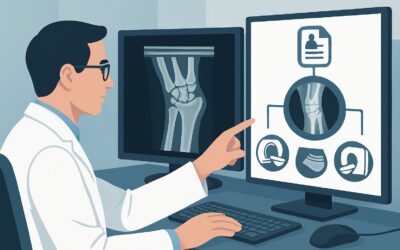
Why Are Radiographs the Recommended First Step for Local Recurrence Surveillance?
For routine surveillance of a primary bone tumor site, the ACR identifies Radiography area of interest as a Usually Appropriate initial study. This recommendation is based on a balance of diagnostic utility, accessibility, and safety.
Radiographs serve as an excellent, low-cost baseline and screening tool. They are highly effective for assessing the bone-hardware interface, detecting new bone destruction (lysis) or formation (sclerosis), and identifying periosteal reactions that may signal recurrent tumor activity. For osteoid-forming tumors like osteosarcoma, radiographs are particularly sensitive to the mineralized matrix of a recurrence. The extremely low radiation dose is a major advantage in a surveillance setting, where patients will undergo many imaging studies over their lifetime.
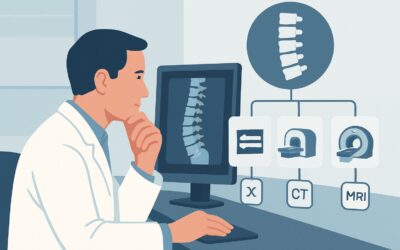
Why are other advanced modalities not the first choice?
- MRI without and with IV contrast is also rated Usually Appropriate but is typically reserved as a second-line or problem-solving tool. While MRI offers superior soft-tissue contrast and is invaluable for assessing non-mineralized tumor components and marrow infiltration, it is more expensive, less accessible, and more susceptible to artifacts from metallic hardware. It is often the next step if radiographs are suspicious or equivocal.
- CT with IV contrast is rated May be appropriate. CT provides excellent detail of cortical bone and can be useful when metallic artifact severely degrades MRI quality. However, its routine use for surveillance is discouraged due to a significantly higher radiation dose compared to radiography and inferior soft-tissue contrast compared to MRI.
- Whole-body bone scan is rated Usually not appropriate for this specific indication. While sensitive for osteoblastic activity, it is nonspecific. Post-surgical inflammation, healing, and hardware loosening can all cause radiotracer uptake, making it difficult to distinguish these benign processes from tumor recurrence.
The strategy of starting with radiographs leverages a high-value, low-risk study to screen for changes, reserving more advanced, higher-cost, or higher-radiation modalities for when they are clinically indicated.
What’s the Next Step After Surveillance Radiographs?
The results of the surveillance radiograph dictate the subsequent clinical workflow. The goal is to confidently rule out recurrence or, if it is suspected, to characterize it and guide a biopsy.
If Radiographs Are Negative or Stable
If the radiographs show no new suspicious findings and are stable compared to prior examinations, the patient can continue their standard surveillance schedule. No further imaging is typically required at this visit. This is the most common and desired outcome.
If Radiographs Are Positive or Suspicious for Recurrence
When radiographs reveal a new lytic or sclerotic lesion, progressive radiolucency at the bone-implant interface, or a new soft tissue opacity, further investigation is mandatory. The next step is almost always MRI of the area of interest without and with IV contrast. MRI will delineate the extent of the suspected recurrence, particularly its soft tissue component, relationship to neurovascular structures, and any intramedullary involvement. These findings are critical for surgical planning and guiding a biopsy.
If Radiographs Are Indeterminate or Equivocal
Post-surgical changes can often make radiographs difficult to interpret. In these cases, where subtle changes are present but not definitively indicative of recurrence, the workflow also proceeds to MRI without and with IV contrast. MRI can differentiate post-operative fibrosis and fluid collections from solid, enhancing recurrent tumor tissue. In select cases with extensive hardware causing prohibitive MRI artifact, a CT scan may be considered as an alternative.
Pitfalls to Avoid (and When to Get Help)
Navigating bone tumor surveillance requires careful attention to detail to avoid common errors that can delay diagnosis or lead to unnecessary procedures.
- Pitfall 1: Neglecting Comparison Films. Interpreting surveillance radiographs in isolation is a major error. The most critical factor is change over time. Always ensure the interpreting radiologist has access to the entire series of prior imaging, especially the initial post-operative baseline.
- Pitfall 2: Misinterpreting Hardware Artifacts. Metallic implants create artifacts on all imaging modalities. On radiographs, “stress shielding” can cause benign bone resorption that can be mistaken for lytic recurrence. Understanding these expected findings is key.
- Pitfall 3: Over-reliance on a Single Modality. While radiographs are the appropriate first step, they have limitations, particularly for detecting non-mineralized soft tissue recurrence. Maintain a low threshold to proceed to MRI if clinical suspicion is high despite negative radiographs.
If imaging findings are equivocal or if there is a discrepancy between clinical suspicion and imaging results, the case should be discussed at a multidisciplinary tumor board including musculoskeletal radiologists, orthopedic oncologists, and pathologists.
Related ACR Topics and Tools
For a comprehensive overview of imaging for all clinical variants related to primary musculoskeletal tumors, and for tools to help with study selection and patient communication, the following resources are available.
- For breadth across all scenarios in Malignant or Aggressive Primary Musculoskeletal Tumor-Staging And Surveillance, see our parent guide: Malignant or Aggressive Primary Musculoskeletal Tumor-Staging And Surveillance: ACR Appropriateness Decoded.
- To explore other clinical situations or find guidance on different presentations, use the ACR Appropriateness Criteria Lookup.
- For detailed technical parameters on how to perform the recommended studies, consult the Imaging Protocol Library.
- To help discuss cumulative radiation exposure with patients undergoing long-term surveillance, the Radiation Dose Calculator can be a useful aid.
Frequently Asked Questions
Why isn’t MRI the first-line study for every surveillance visit?
While MRI is excellent for soft tissue and marrow evaluation, it is more costly, time-consuming, and susceptible to metallic artifact than radiography. For routine surveillance in an asymptomatic patient, radiographs provide a highly effective, low-radiation, and cost-effective screening tool. MRI is reserved for problem-solving when radiographs are suspicious or clinical concern is high.
How often should surveillance radiographs be performed?
The frequency of surveillance imaging depends on the specific tumor type, its grade, the time since treatment, and institutional or cooperative group protocols. Generally, surveillance is more frequent in the first 2-3 years after treatment, when the risk of recurrence is highest, and then spaced out over time.
What if my patient has a large endoprosthesis that causes significant artifact?
Extensive metallic hardware is a known challenge. While modern MRI techniques like metal artifact reduction sequences (MARS) can significantly improve image quality, severe artifact can still be limiting. In such cases, CT may be a more useful problem-solving tool than MRI. FDG-PET/CT, rated ‘May be appropriate,’ can also be valuable in complex post-surgical cases to assess for metabolically active recurrence.
Does the type of original bone tumor (e.g., osteosarcoma vs. Ewing sarcoma) change this recommendation?
For the initial step in local surveillance, the recommendation to start with radiographs is generally consistent across the common primary malignant bone tumors. However, the specific appearance of recurrence and the relative utility of subsequent imaging (like MRI vs. PET/CT) can vary. For example, radiographs are particularly useful for detecting the mineralized matrix of recurrent osteosarcoma.
Should I order a whole-body bone scan for surveillance?
No, for surveillance of *local recurrence*, a whole-body bone scan is rated ‘Usually not appropriate’ by the ACR. Its low specificity is a major drawback, as post-surgical changes, inflammation, or hardware issues can all cause tracer uptake, leading to false-positive results that trigger unnecessary workups.
Reviewed by Pouyan Golshani, MD, Interventional Radiologist — May 30, 2026